LESSON ASSIGNMENT
LESSON 10 Cardiovascular and Other Circulatory Systems of the Human Body.
LESSON ASSIGNMENT Paragraphs 10-1 through 10-45.
LESSON OBJECTIVE After completing this lesson, you should be able to identify functions of the cardiovascular and lymphatic systems; including functions of their parts.
SUGGESTION After completing the assignment, complete the exercises at the end of this lesson. These exercises will help you to achieve the lesson objectives.
LESSON 10
CARDIOVASCULAR AND OTHER CIRCULATORY SYSTEMS OF THE
HUMAN BODY
Section I. INTRODUCTION
10-1. NEED FOR A CIRCULATORY SYSTEM
In simple organisms such as unicellular and one-or two-layer organisms, materials can be transferred among cells by simple processes of diffusion. However, in large organisms, a system is needed for the distribution and collection of materials. This is because diffusion does not occur fast enough to carry the large volumes of materials necessary through the greater distances required.
10-2. DISTRIBUTION OF SUBSTANCES
a. Products of the Digestive System. Some of the substances distributed to the body cells are products of the digestive system. These materials meet individual cell requirements for energy, growth, repair, synthesis of new materials, and storage for later use.
b. Oxygen. In the lungs, oxygen is obtained by the blood through the process of external respiration. Oxygen is then transported to the individual body cells, where it is used in metabolic oxidation. This provides energy for production of ATP (adenosine triphosphate), which is necessary for carrying on the life processes of the body.
10-3. COLLECTION OF SUBSTANCES
Some substances are collected from the body cells for elimination. These include carbon dioxide, nitrogenous wastes, and other potentially harmful substances that are carried to organs like the lungs, liver, or kidneys for elimination from the body.
10-4. HORMONES AND OTHER CONTROL SUBSTANCES
Hormones are the products of endocrine glands (see lesson 11). Hormones and other control substances are distributed throughout the body by circulatory systems. The tissues or organs affected by these substances are usually called target organs. In turn, substances released by the target organs often affect the original endocrine gland. This results in a feedback system.
10-5. CONTINUOUS RENEWAL AND REMOVAL OF FLUIDS
Secretory processes continuously renew the various fluid systems of the human body. At the same time, the volume of fluid in each system is kept at a constant level through the removal of excess fluids. Should the removal processes be interrupted, the volume of fluid will increase. The resulting increase in pressure can have serious consequences. Depending on the system involved, the consequences might include deafness, hydrocephalus, or pulmonary edema.
10-6. COMPONENTS OF ANY CIRCULATORY SYSTEM
Any circulatory system has three general components:
a. Vehicle. The vehicle is a fluid (flowing) medium. The materials being carried are dissolved or suspended in this fluid. This is the blood, lymph, or cerebrospinal fluid.
b. Conduits. Conduits are like pipes. They contain the fluids in which materials are transported to and from the various parts of the body. These are the blood vessels or lymph vessels.
c. Motive Forces. Motive forces act upon the vehicle to make it flow through the conduits. These are provided by the heart.
10-7. EXAMPLES OF CIRCULATORY SYSTEMS
Some circulatory systems of the human body are the cardio-vascular system, the lymphatic system, and the CSF (cerebrospinal fluid) system. The lesser systems include the aqueous humor of the bulbus oculi (eyeball) and the endolymph and perilymph, which are fluids of the inner ear.
10-8. INTRODUCTION TO THE CARDIOVASCULAR SYSTEM
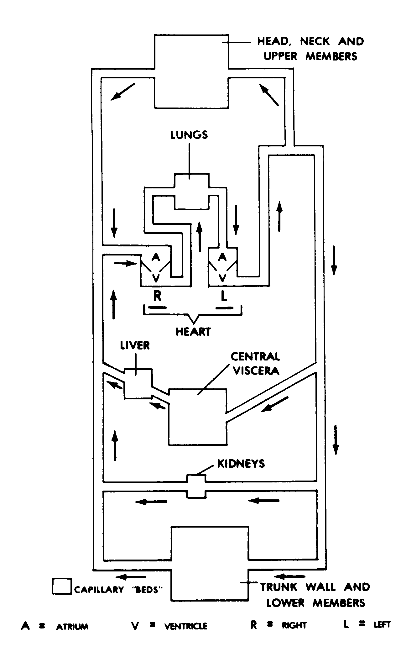
The cardiovascular system (Figure 10-1) is the primary circulatory system of the human body. It includes a heart, blood, and blood vessels.
a. One function of the cardiovascular system is transport. Some substances carried by the cardiovascular system are dissolved or suspended in the fluid portion of the blood. Others are bound up in special cellular elements (RBCs).
b. The cardiovascular system also provides protection against foreign substances. This function involves active attack by white blood cells as well as more subtle processes of the immune system.
Figure 10-1. Diagram of the human cardiovascular (cirulatory) system.
Section II. THE BLOOD--THE VEHICLE OF THE CARDIOVASCULAR SYSTEM
10-9. DEFINITION
Blood is the vehicle of the cardiovascular system. Thus, the component actually transports substances.
10-10. PLASMA
Plasma makes up about 55 percent of the total blood volume.
a. Water. The major constituent of plasma is water. The physical characteristics of water make it a very good vehicle.
(1) Since water is fluid, it can flow through the conduits.
(2) Since most substances can be dissolved in water, it is often known as the "universal solvent."
(3) At ordinary pressures, water is essentially non-compressible.
(4) In addition, water has important temperature characteristics.
(a) Water has an ample heat-carrying capacity. It can carry heat readily throughout the body.
(b) Some of this heat is transferred to the water of the sweat glands. Since water can dissipate great quantities of heat through evaporation, excess heat can be efficiently disposed of at the surface of the skin.
b. Dissolved and Suspended Substances. To some extent, all transported substances are dissolved or suspended in the water of the plasma. These substances include various gases, end products of digestion, various control substances, and waste products. Also, there are three major plasma proteins--albumin, globulins, and fibrinogen. Together with dissolved salts (electrolytes), these plasma proteins help to maintain the tonicity of the plasma. In addition, fibrinogen is important to blood clotting.
10-11. FORMED ELEMENTS
The remainder of the blood volume consists of the formed elements--the red blood cells, the white blood cells, and the platelets. In adults, these formed elements normally make up 40 percent to 45 percent of the total blood volume. (This measure is called the hematocrit.)
a. Red Blood Cells (RBCs; Erythrocytes). The primary function of RBCs is to contain the protein called hemoglobin, which in turn carries oxygen. Thus, RBCs carry the majority of the oxygen to the individual cells of the body.
(1) Structure. The normal, mature red blood cell is a biconcave disc. The biconcave shape results from the loss of the nucleus just before the final maturation of the RBC. Since this shape increases the surface area of the disc, there is an increase in the capacity for the flow of substances into and out of the RBC.
(2) Hemoglobin. Within the cytoplasm of the RBC is a special protein called hemoglobin. Because of its iron atoms, hemoglobin has a great affinity for oxygen. It will readily pick up oxygen until it is saturated. At the same time, however, hemoglobin will readily give up oxygen in areas of low concentration.
(3) Life cycle of the RBC. Because of the loss of its nucleus, the RBC has a limited life period (about 120 days). At the end of this period, the spleen removes the "worn out" RBC, and the liver salvages the "pieces," particularly the iron.
b. White Blood Cells (WBCs; Leukocytes). The white blood cells are also formed elements of the blood. There are several types.
(1) Neutrophils and other phagocytic WBCs. The phagocytic WBCs can move independently out of the capillaries and penetrate into the tissues of the body. There, they actively attack foreign substances and engulf them in a process called phagocytosis. When these WBCs are overcome by foreign substances and die, their bodies accumulate to form a substance called pus.
(2) Lymphocytes. The lymphocytes are involved with the immune system of the body, including the production of antibodies.
c. Platelets. The platelets are the third type of formed element in the blood. Platelets are fragments of former cells. They are very important in the clotting process.
10-12. SERUM
After blood has been treated to remove the formed elements and the protein fibrinogen, there is a clear light-straw-colored fluid remaining. This fluid is called serum.
10-13. TRANSPORT OF GASES
One very important transport function of the blood is to carry gases back and forth between the lungs and the individual cells of the body. The alveoli and the individual body cells are the sites of exchange of gases to and from the blood. At these sites, the gases move according to the directions of pressure of concentration gradients. That is, each gas moves from an area where it is in higher concentration to an area of lesser concentration.
a. Oxygen. Oxygen is in the air filling the alveolus of the lung. The oxygen passes through the walls of the alveolus and capillary to become dissolved in the plasma of the blood. Most of the dissolved oxygen is rapidly picked up by the hemoglobin of the RBCs. Thus, the RBC is the main transporting element for oxygen in the blood.
b. Carbon Dioxide. Carbon dioxide is produced during metabolic oxidation within the individual cell. It passes through the cell membrane and the wall of the capillary to become dissolved in the plasma of the blood. Through action of an enzyme in the RBCs, most of the carbon dioxide (CO2) is transformed into bicarbonate ions (HCO3).
10-14. TRANSPORT OF OTHER SUBSTANCES
Other substances, such as the end products of digestion, are also carried by the blood. They are either dissolved or suspended in the plasma.
10-15. IMPORTANCE OF BLOOD IN ENERGY MOBILIZATION
The life processes cannot continue in the body cells without sources of energy. From glucose, energy is released to produce ATP, the driving force of the life processes of the body.
a. When a specific portion of the cerebral cortex is active, more blood is delivered to that portion. This is an example of how more blood can be delivered to the body parts where it is most needed.
b. When the hormone epinephrine (Adrenalin) is secreted by the adrenal gland, it is delivered to all parts of the body by the cardiovascular system. Among other effects, epinephrine increases the rate of metabolism of all cells of the body. This helps to mobilize energy during a "fight-or-flight" stress reaction.
c. In periods when much energy is required, the body can use its stores of fat as a source of energy. As we have seen in the chapter on the digestive system, the lymphatic circulatory system picks up the end products of lipid (fat) digestion and carries them to the cardiovascular system.
(1) This fat is generally deposited throughout the body, particularly the subcutaneous layer, as yellow fat. In a rapid turnover, the high energy content of the fat is released for use throughout the body.
(2) In infants, there is often brown fat at the junctions of the major blood vessels. In periods of high-energy requirements, this brown fat releases energy into the blood stream immediately.
10-16. RESPONSES TO HEMORRHAGE
A blood vessel may be damaged by transection (cutting across) or rupture. At such points, a volume of whole blood can flow out of the blood vessels. This escape of blood from the blood vessels is called hemorrhage.
HEMO = blood
RRHAGE = excessive flow ("bursting forth")
a. Vascular contraction. The first response to a cut or ruptured vessels is contraction (spasm) of the blood vessel itself. This may considerably reduce the volume of blood loss.
b. Platelet Plug. If the hole is small, a plug formed by clumping of the platelets may be adequate to stop the bleeding.
c. Blood Clotting. There is a complicated process for sealing off holes or ends of blood vessels after a cut or rupture. By this process, called coagulation or clotting, the blood forms a solid mass to seal the opening where the blood is escaping. The mass is called a blood clot. After many intermediate steps, the protein fibrinogen of the blood is converted into sticky strands of fibrin. These sticky strands adhere to the wall of the opening and form a meshwork in the opening, which traps RBCs and plasma. Thus, the opening is sealed.
d. Hematoma. A hematoma is a collection of blood, usually clotted, in an organ, space, or tissue. When found immediately beneath the skin, it will produce a purplish spot or mark. With time, as the clot is broken down and resorbed, the hematoma changes color and becomes smaller.
e. Mobilization of Blood Reservoirs. Certain areas of the body contain enough blood that they can be used as reservoirs to maintain the circulating blood volume. This is important when a volume of blood has been lost through hemorrhage. Among these are the spleen and the liver, whose sinuses together can release several hundred milliliters of blood. Also important are several groups of veins, including the large abdominal veins, which can also provide several hundred milliliters of blood.
10-17. BLOOD TRANSFUSIONS AND BLOOD MATCHING
a. Transfusions. In cases where an individual has lost whole blood by hemorrhaging, it is often necessary to give transfusions of whole blood. Whole blood transfusions continue the functions of the RBCs. On the other hand, if an individual has suffered burns causing a loss of fluid but not the loss of formed elements, plasma or a plasma substitute will often be used.
b. Blood Matching. There are a number of substances (antigens) on the surfaces of RBCs that vary among individuals. The blood of other individuals may contain or develop antibodies to these antigens. Before blood transfusions, the blood of the recipient and the donor must be matched to avoid potentially fatal reactions. Important systems of such antigens include the ABO system and the Rh system.
Section III. THE BLOOD VESSELS--THE CONDUITS OF THE CARDIOVASCULAR SYSTEM
10-18. INTRODUCTION
The blood vessels are tubular structures throughout the entire body. Since this tubular system is continuous (without interruption or opening), we sometimes refer to it as a closed system.
10-19. TYPES OF BLOOD VESSELS AND THEIR CONSTRUCTION
In general, there are three types of blood vessels--arteries, veins, and capillaries. We use the following abbreviations:
A. = artery V. = vein
Aa. = arteries Vv. = veins
NAVL = nerve(s), artery(ies), vein(s), lymphatic(s)
a. Three General Layers. In general, a blood vessels has a wall composed of three layers.
(1) Intima. The innermost layer is the intima. The intima is a simple epithelium made up of a single layer of flat epithelial cells.
(2) Media. The main portion of the wall is the media. It is made up of a combination of FCT and smooth muscle tissue.
(3) Adventitia. The outer surface of the blood vessel is the adventitia. It is an FCT layer.
b. Comparison of the Structures of Arteries and Veins. Given an artery and a vein with similar inner diameters, the artery will have a thicker wall than the vein. This greater thickness is due to the presence of more smooth muscle tissue and the presence of elastic FCT as a significant element.
c. Capillary Structure. Capillary walls have only one layer--the intima. Capillary networks (beds) are the exchange areas for the cardiovascular system. This includes the internal exchange areas between the blood and the individual cells of the body. Since the capillary wall consists of flat single cells, substances can move readily between the body cells and the blood.
10-20. SPECIAL SITUATIONS
This paragraph describes several special situations associated with the blood vascular system.
a. Nutrient Versus Functional Blood Supplies. The lungs, liver, and heart actually have two blood supplies. The functional blood supply provides blood to be worked upon by the organ. The nutrient blood supply provides blood for the usual exchange of materials between body cells and the blood.
b. Collateral Circulation. A collateral circulation is a special organization of blood vessels around a major joint of other area of the body. Its purpose is to provide a continuing supply of blood even if one of the vessels is damaged. Several blood vessels are included so that there will be an alternate route when needed.
c. End Arteries. There are other areas of the body where a single artery is the sole supply of blood. Such an artery is called an end artery. When an end artery is damaged and can no longer supply blood to an area, the tissues of the areas will die. End arteries are most common in the brain and the heart.
d. Portal Veins. A portal vein is a venous blood vessel that begins with capillaries in one area and ends in capillaries of another area. The most important portal vein in the human body is the hepatic portal vein. The hepatic portal vein extends from the capillaries of the digestive system to the capillaries/sinusoids of the liver.
10-21. LOCATIONS OF BLOOD VESSELS TYPES
In the human body, blood vessels are located differently according to their types.
a. Arteries. If an artery is injured, the threat to life is greater than with other types of blood vessels. For protection, arteries tend to be located deep within the structures of the body. Only the very smallest of arteries, especially the cutaneous arteries, come close to the surface of the body.
b. Veins. There are both deep veins and cutaneous veins. The deep veins accompany the arteries side by side. The cutaneous veins are found in the subcutaneous layer of the body. The cutaneous veins drain into the deep veins at specific locations (especially the inguinal region and the axillary region).
c. Capillaries. The capillaries are located throughout all tissues of the body. No individual cell is more than two cells away from a capillary. The networks of capillaries in the tissues are often called capillary beds.
10-22. PATTERNS OF BLOOD CIRCULATION
Blood vessels make up a closed system, since there is no place in the system where whole blood can leave.
a. Direction of Flow of Arteries and Veins. Arteries carry blood from the chambers of the heart to the tissue of the body. Veins carry blood from the tissues to the chambers of the heart. (Coronary arteries carry blood from the chambers of the heart inside to the walls of the heart outside.)
b. Two-Cycle System. It is also a two-cycle system (Figure 10-2). It involves both the pulmonary cycle and the systemic cycle. Blood circulates through two circuits. In the pulmonary cycle, blood circulates from the heart to the lungs and back to the heart. In the systemic cycle, blood circulates from the heart to the rest of the body and back to the heart.
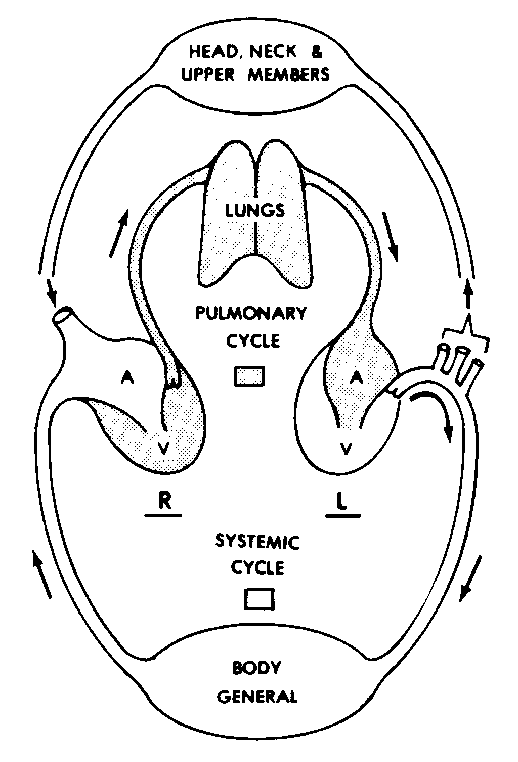
Figure 10-2. Cardiovascular circulatory pattern.
c. Fetal Circulation. Since the fetus is located within the uterus, its lungs do not take in air. Therefore, the pulmonary cycle does not function in the fetus. Essentially, fetal blood flows to and from the placenta. There are certain bypasses in the heart to avoid the pulmonary cycle. At the time of birth, the fetal circulation is changed to the normal pattern.
Section IV. THE HEART--THE PRIMARY MOTIVE FORCE OF THE CARDIOVASCULAR SYSTEM
10-23. INTRODUCTION
In humans, the heart is the primary motive force for driving the blood along the arterial vessels. The heart consists of four separate chambers. Two chambers function as a "right heart," and two function as a "left heart." The muscular walls (myocardium) of the chambers apply force to the blood within and force the blood to move out of the chambers. (See Figure 10-3.)
10-24. CHAMBERS OF THE HUMAN HEART
a. Atria. Two chambers are called the atria (singular: atrium). Down the middle, an interatrial septum separates the two atria.
(1) The muscular walls of the atria tend to be relatively thin.
(2) Attached to each atrium is an earlike appendage called an auricle. The auricles of the atria tend to have somewhat thicker walls.
b. Ventricles. The other two chambers are the right and left ventricles. Between the ventricles is the interventricular septum.
(1) The left ventricle tends to be cylindrical in shape. It has a relatively thick wall.
(2) The right ventricle has a somewhat semilunar (half-moon) cross section, since it is wrapped around one side of the left ventricle.
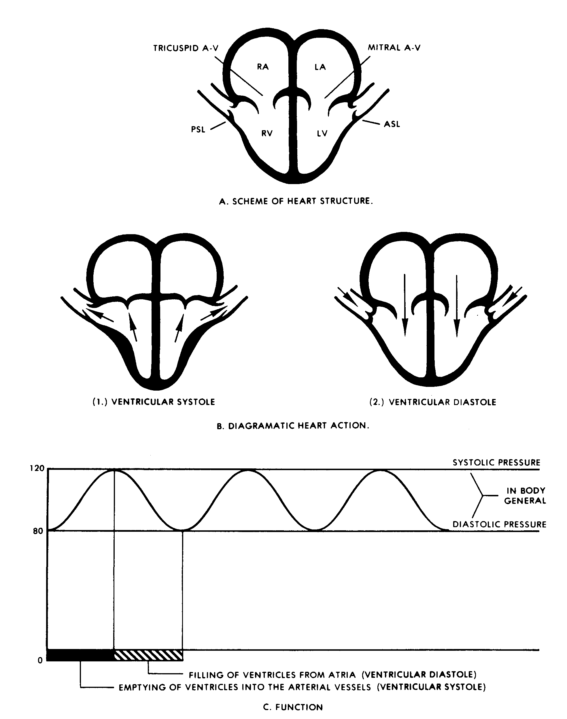
Figure 10-3. The human heart function.
10-25. FIBROUS SKELETON OF THE HEART
There is an FCT structure within the substance of the heart. This structure is known as the fibrous skeleton of the heart. This fibrous skeleton serves two general purposes: (1) as sites of attachment for muscle tissues and (2) as supporting structures for the cardia valves. All of the fibrous structures are continuous and form the fibrous skeleton of the heart.
a. Fibrous Portion of the Interventricular Septum. The uppermost portion (also called the membranous portion) of the interventricular septum is a part of the fibrous skeleton of the heart.
b. Atrioventricular (AV) Rings. Each atrioventricular valve of the heart is surrounded by a dense fibrous ring. This ring maintains the valve opening.
c. Cylinders at Bases of Great Arteries. Each of the semilunar valves of the heart is located within a short fibrous cylinder. This cylinder maintains the structure and function of the valve.
10-26. WALL STRUCTURE
The walls of the chambers of the heart are in three layers.
a. The chambers themselves are lined with a simple epithelium known as the endocardium.
b. Likewise, a simple epithelium surrounds the outside of the heart. It is known as the epicardium. The epicardium is the same as the visceral pericardium, which we shall discuss later.
c. By far the most important is the myocardium, the middle layer. It is made up of cardiac muscle tissue.
(1) Cardiac muscle tissue consists of fibers formed by the fusion of many individual cells (syncytium). These cardiac fibers are striated and branched.
(2) The myocardium is thicker in the walls of the ventricles than the atria. This is because greater pressures are needed for the ventricles to perform their function. The wall of the left ventricle is especially thick, since it has to drive the blood throughout the body.
(3) The inner surfaces of the ventricular walls have ridges of muscle known as the trabeculae carneae, with spaces between them.
(4) When the musculature within a chamber wall contracts, the lumen (cavity) decreases in diameter. This is particularly true of the left ventricle. There is also a twisting or wringing action of the left ventricle that causes the apex of the heart to hit against the inner surface of the chest wall--the apex beat.
(5) The stroke volume is the amount of blood forced out of each ventricle in one contraction. The cardiac output is the volume of blood pumped out of the ventricles (RT into the lungs, LT into the systemic circulation) in one minute (expressed in liters per minute). These volumes will change according to the needs of the body.
10-27. CARDIAC VALVES
Valves are structures that ensure that fluids will pass through them in only one direction. That is, a valve will open to allow fluids to pass in one direction but will close to prevent fluids from passing in the other direction. There are two sets of cardiac valves--the atrioventricular (AV) valves and the semilunar valves. Although the two sets of valves are quite different in design, they both function passively in response to the flow of the blood.
a. AV Valves. The AV valves are found between the atria and the ventricles. The AV valves consist of flaps, known as cusps. The outer margin of each flap is attached to the inner surface of a fibrous ring. The inner edge of each flap is free.
(1) On the right side is the tricuspid valve. On the left side is the mitral valve. ("Might is never right.")
(a) Thus, the tricuspid valve is between the right atrium and the right ventricle. It is named for its three cusps.
(b) The mitral valve is located between the left atrium and the left ventricle. Since it has two cusps, it is sometimes called the "bicuspid" valve.
(2) The contraction of the atrial walls forces the blood from the atria through the AV valves and into the ventricles (atrial systole).
(3) When the atria relax (atrial diastole) and the ventricles contract, the pressure would tend to drive the blood back into the atria. However, each opening is sealed when the cusps of each AV valve meet in the valve center. This prevents blood from flowing further back into the atria.
(4) A special anatomic arrangement helps prevent backward flow into the atria. Chordae tendineae are fibrous cords attached to the ventricular side of the cusps. Since these cords of dense FCT have a fixed length, they cannot be stretched or shortened. The other ends of these cords are attached to the papillary muscles. The papillary muscles are special extensions of the muscular walls of the ventricles. As the ventricles contract and become smaller, these muscles take up the slack in the cords.
b. Semilunar (Aortic and Pulmonary) Valves. As mentioned before, the bases of the two great arteries (the pulmonary arch and the aortic arch) begin at their respective ventricles as short cylinders of the fibrous skeleton. Within each of these cylinders are three cuplike cusps, which make up each semilunar valve. When the ventricles contract (ventricular systole) and the AV valves have closed, the blood moves out into the great arteries through the semilunar valves. When the ventricles relax (ventricular diastole), the back pressure of the blood in the great arteries forces the cusps of the semilunar valves to the center and seals off each opening.
10-28. NAVL OF THE HEART
a. Controls of Heart Function.
(1) Extrinsic controls. A number of cardiac nerves arise from both the sympathetic and parasympathetic portions of the nervous system (chapter 12). The sympathetic portion accelerates the action of the heart, while the parasympathetic portion slows it down. These portions are both controlled by cardiovascular centers in the medulla of the hind-brainstem. In addition, as everyone is well aware, various emotional states can affect the actions of the heart.
(2) Intrinsic controls. Within the substance of the heart, certain fibers of the myocardium have been transformed from contracting muscle tissue to impulse-transmitting fibers. These are called Purkinje's fibers. Together, these fibers provide intrinsic control for the action of the heart.
(a) The sinoatrial (SA) node is a collection of these fibers in the interatrial septum. The SA node is often called the pacemaker of the heart because it initiates each cycle of the contractions of the heart chambers.
(b) The atrioventricular (AV) node is another group of these fibers just above the interventricular septum.
(c) Descending from the AV node is the bundle of His, which branches into the right and left septal bundles. These branches pass down on either side of the interventricular septum.
(d) Impulse begin in the SA node, pass to the AV node, and then descend through the septal bundles to stimulate the myocardium of the ventricular walls to contract.
(3) Humoral control. Apparently, some substances transported by the blood can accelerate or slow the action of the heart. This situation is called the humoral control of heart action.
b. Coronary Arteries. Previously, we have described the flow of blood through the chambers of the heart. This blood, upon which the heart acts, is called functional blood. Now, we wish to discuss the supply of nutrient blood to the heart. This blood nourishes the tissues of the heart. The nutrient blood supplies oxygen and food materials to the tissues of the heart and removes waste materials. This nutrient blood is supplied to the walls of the heart by the right and left coronary arteries.
(1) The openings leading into the coronary arteries are located in the base of the ascending aorta, just above (behind the cusps of) the semilunar valve (aortic valve). When this valve is open, its cusps cover the openings of the coronary arteries. When the valve is closed, the backpressure of the blood in the aorta fills the coronary arteries with blood. The coronary arteries then distribute the blood to all of the tissues of the relaxed heart.
(2) Many of the branches of the coronary arteries are of the end artery type. This means that such a branch is the sole supply of nutrient blood to a specific area of the heart. If the branch should be closed for any reason, the tissue in that area will die for lack of oxygen and nourishment.
c. Cardiac Veins and Coronary Sinus. The blood from the tissues of the heart is collected by the cardiac veins. These veins empty into the coronary sinus, a vessel, which in turn empties into the right atrium.
d. Thebesian Veins. The thebesian veins are many minute sinuses found in the myocardium of the ventricles. They extend from the lumen into the myocardium of each ventricle.
10-29. HEART SOUNDS
When the valves of the heart close, they produce audible sounds. First, the closing of the AV valves produces a noticeable "LUB." When the semilunar valves subsequently close, another sound "DUB" is produced to complete the cycle. These are referred to as the heart sounds--"LUB DUB, LUB DUB," etc.
10-30. ELECTROCARDIOGRAM (EKG)
Since the myocardial tissue is living material, its activity produces electrical impulses. With an electrocardiogram, the pattern of these electrical impulses can be recorded.
10-31. THE PERICARDIUM
a. General. The heart is an active organ of the human body. Its pumping action, which begins in the very early embryo, continues without stopping until death. During each cycle of its activity, the heart changes in shape and size and tends also to rotate. (The number of cycles per minute is called heart rate.) To reduce the amount of friction resulting from this activity, the heart is includes within a serous sac, called the pericardium, or pericardial sac.
b. Serous Space and Two Serous Pericardia. As in all serous cavities, there is a serous space between two serous membranes.
(1) The visceral pericardium intimately covers the surface of the heart. Earlier, we referred to this as the epicardium.
(2) The parietal pericardium is the outer serous membrane.
(3) Between the two serous pericardia is a very thin space containing a thin film of pericardial fluid. This lubricating fluid makes the action of the heart much less strenuous.
c. Fibrous Pericardium. The parietal pericardium is covered with a very dense fibrous envelope. This envelope forms the outer portion of the pericardial sac.
Section V. MOTIVE FORCES INVOLVED IN DRIVING THE BLOOD THROUGH THE SYSTEM
10-32. INTRODUCTION
The blood (vehicle for transporting material) is driven through the blood vessels (conduits) by a variety of motive forces.
10-33. ARTERIAL BLOOD FLOW
Blood is driven through the arteries by a combination of forces. First, there is the force produced by the contraction of the ventricular walls. Second, there is the elastic recoil of the arterial walls.
a. Systole. When the left ventricle contracts (systole), it forces the blood into the aortic arch. Above the base cylinder, the wall of the aortic arch is mainly elastic FCT. As the blood fills the aortic arch, the walls are stretched.
b. Diastole. When the ventricle relaxes (diastole), the wall of the arch recoils and presses against the blood. With the closing of the aortic semilunar valve, the blood is forced to move out along the arteries in a pressure pulse. Since the elasticity of the arterial walls produces a continuous pressure, the blood moves continuously throughout the system.
c. Arterial Pressures. The highest pressure is called the systolic pressure, and the lowest pressure is the diastolic pressure.
d. Vasoconstriction. Vasoconstriction is the actual contraction of the arterial walls. Vasoconstriction can further increase the pressure on the blood in the arteries.
e. Gravity. Gravity helps to move blood to the trunk and lower members. However, it is a hindrance in moving blood to the head and neck.
10-34. VENOUS BLOOD FLOW
There is usually a low level of pressure in the veins. There are valves in the veins that ensure that blood flows continuously toward the heart. Therefore, as pressure is applied to a vein, there will be a pump effect.
a. Pressure from Arteries. The muscular compartments of the upper and lower limbs tend to be full in healthy persons. Therefore, as blood enters the arteries within these compartments, a volume of blood must leave through the veins.
b. Pressure from Muscular Contractions. During muscular activity, additional forces press against the veins and produce a "milking action." Again, blood moves through the veins back toward the heart.
c. Gravity. In the head and neck, gravity helps to move the blood down through the veins. In the trunk and lower limbs, the valves help to prevent a backward flow of blood in the veins.
Section VI. CAPILLARIES
10-35. INTRODUCTION
The capillary beds make up the greatest cross-sectional area of the cardiovascular system. In the capillary beds, the actual exchange of materials takes place between the blood and the cells of the body.
10-36. FILTRATION PHENOMENON
The wall of the capillary consists of a single layer of flat cells. The minute spaces surrounding the capillaries and the individual cells of the body make up the tissue space (interstitial/ extracellular space). Fluid passes from the capillary into the tissue space and carries with it various substances. Some of this fluid returns to the capillary on the venous side.
10-37. CAPILLARY SPHINCTERS
The capillary beds are provided with precapillary sphincters that can reduce or completely stop the flow of blood into the capillaries. At the other end of the capillary bed are postcapillary sphincters; when these close, there is a backpressure and more fluid flows into the tissue space.
Section VII. TEMPERATURE CONTROL BY MEANS OF THE BLOOD
10-38. ELIMINATION OF EXCESS HEAT
Heat is produced as a by-product by various activities of the human body, particularly muscular contractions. When excess heat is accumulated, it must be eliminated from the body to maintain a healthy condition.
a. The water of the blood has a great heat-carrying capacity.
b. There are superficial capillary beds in the subcutaneous layer, close to the surface of the body. When the blood flows through these beds, some of its heat can radiate directly to the surrounding environment.
c. The sweat glands take water from the blood and secrete it onto the surface of the skin. Here, even more calories of heat are lost during the evaporation of the water.
10-39. CONSERVATION OF BODY HEAT
On the other hand, if the body has an insufficient amount of heat, heat loss must be reduced. For this purpose, the superficial capillary beds can be closed down. Then, the fat in the subcutaneous layer serves as insulation.
10-40. CORE TEMPERATURE CONTROL
Unlike the peripheral portions of the body, whose temperatures may vary considerably, the center of the body must be maintained at a certain temperature within very narrow limits.
a. Control. There are special temperature detectors in the hypothalamus of the forebrainstem. These continuously monitor the temperature of the blood flowing through the brain.
b. Counter-Current Mechanism. The peripheral blood in the limbs is several degrees cooler than the blood in the center of the body. Therefore, it must be warmed as it returns toward the heart. As previously described, the arteries and veins of the limbs are located side by side as they extend from the trunk and through the length of the limbs. As it returns to the trunk, cool venous blood is gradually warmed by the arterial blood flowing in the opposite direction.
10-41. COOLING OF ORGANS WITH A HIGH METABOLIC RATE
Certain organs of the body, such as the brain and the liver, have a relatively high metabolic rate. Because of this, they produce excessive heat. Part of the blood supply to these organs is specifically designed to remove the excess calories of heat.
10-42. WARMING OF INFLOWING AIR
As blood flows through the arteries of the mucoperiosteum of the nasal chambers, the inflowing air is warmed.
10-43. ERYTHEMA
At the site of an infection or injury, the most common reaction observed is redness (erythema). This indicates that extra blood and heat are available for healing.
Section VIII. OTHER CIRCULATORY SYSTEMS
10-44. THE LYMPHATIC SYSTEM
In general, the lymphatic system is a drainage system that picks up tissue fluids and returns them to the cardiovascular system. The tissue fluids are picked up in the interstitial spaces. They are eventually returned to the veins.
a. Lymphatic Capillaries and Vessels.
(1) Within the tissue spaces, the lymphatic system begins with lymph capillaries. A lymph capillary begins with a blind end (cul-de-sac).
(2) The capillaries eventually come together to form lymphatic vessels, which gradually join and become larger and larger. Physiologically, lymphatic vessels are very similar to veins. Like veins, they have low pressure and possess valves.
(3) The thoracic duct is a major collecting vessel of the lymphatic system that empties into the deep veins of the neck. It begins in the upper posterior abdomen with a collection of sacs called the cisterna chyli. The cisterna chyli is a receiving area for lymph from three other major lymphatic vessels. From the cisterna chyli, the thoracic duct passes upward through the thorax and into the root of the neck. There, it empties into the deep veins of the neck.
b. Lymph Nodes. Along the lymphatic vessels at various intervals are small structures known as lymph nodes. The lymph nodes function as sieves for the lymph passing through them. In healthy individuals, the lymph nodes usually draw no attention. However, in chronic diseases, the lymph nodes become enlarged and hardened (indurated). In the axilla, the inguinal region, and the neck, certain lymph nodes are large enough to be palpated even in health. Tonsils are aggregates of lymphatic tissue.
c. Lymphocytes. Associated with the lymphatic system are special cells known as lymphocytes. The lymphocytes become part of the formed elements of the blood. They are primarily involved in the immune reactions of the body.
10-45. CIRCULATORY SYSTEMS OF LESSER VOLUME
In addition to the cardiovascular and lymphatic circulatory systems, there are other circulatory systems of lesser volume.
a. The cerebrospinal fluid (CSF) system is involved with the central nervous system. CSF is formed with fluid from the arteries and eventually returned to venous vessels.
b. The bulbus oculi (eyeball) and the inner ear are fluid-filled hollow organs. Such organs have their own internal circulatory systems. In the case of the bulbus oculi, the fluid is the aqueous humor. In the case of the inner ear, the fluid is the endolymph/perilymph. In such cases, the fluids are produced from fluids of arterial vessels and then are picked up by venous vessels. Should the drainage pattern be interrupted, fluids will accumulate and cause increased pressure within the hollow organ. The increased pressure will interfere with the organ functions; examples are glaucoma of the eye and deafness of the ear.