LESSON ASSIGNMENT
LESSON 12 The Human Nervous System.
LESSON ASSIGNMENT Paragraphs 12-1 through 12-38.
LESSON OBJECTIVES After completing this lesson, you should be able to:
12-1. Identify the major subdivisions of the human nervous system.
12-2. Match terms related to the human nervous system with their definitions.
12-3. Identify body functions and classes of organs and tissues which are the concern of major subdivisions of the human nervous system.
12-4. Given a list of statements about one of the following topics, identify the false statement.
a. Electrochemical transmission of neuron impulses.
b. General sensory and motor pathways.
c. Levels of control in the human nervous system.
SUGGESTION After completing the assignment, complete the exercises at the end of this lesson. These exercises will help you to achieve the lesson objectives.
LESSON 12
THE HUMAN NERVOUS SYSTEM
Section I. INTRODUCTION
12-1. THE NEURON
The neuron (nerve cell) is the conducting unit of the nervous system. It is specialized to be irritable and transmit signals, or impulses. The neurons are held together and supported by another nervous tissue known as neuroglia, or simply glia.
12-2. MAJOR SUBDIVISIONS OF THE NERVOUS SYSTEM
The human nervous system can be considered in three major subdivisions:
a. The central nervous system (CNS).
b. The peripheral nervous system (PNS).
c. The autonomic nervous system (ANS).
12-3. DEFINITIONS
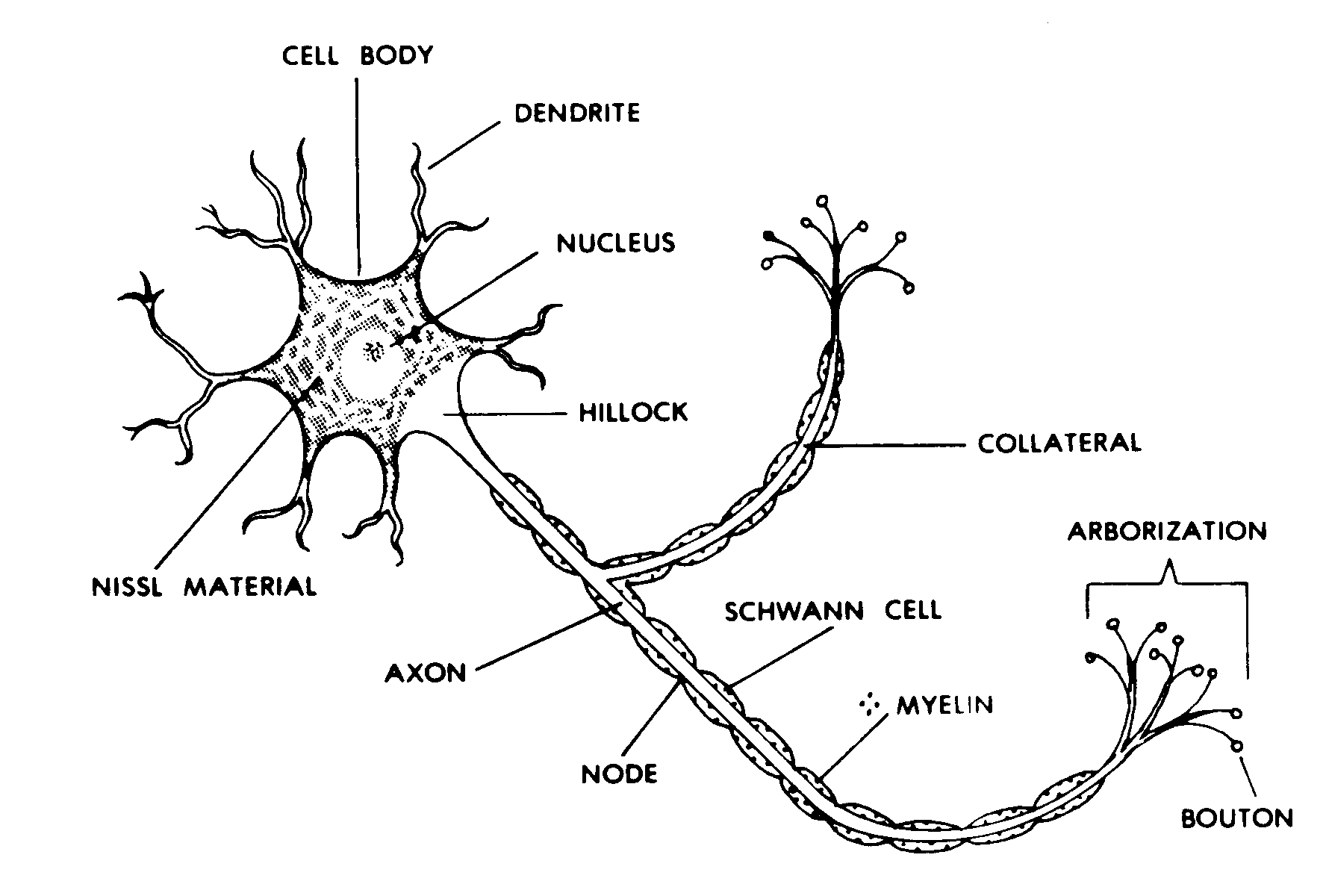
a. Neuron. A neuron (Figure 12-1) is the nerve cell body plus all of its processes and coverings.
Figure 12-1. A "typical" neuron.
b. Nerve. A nerve is a collection of neuron processes together and outside of the CNS.
c. Fiber Tract. A fiber tract is a collection of neuron processes together and within the CNS.
d. Ganglion. A ganglion is a collection of nerve cell bodies together and outside of the CNS.
e. Nucleus. A nucleus is a collection of nerve cell bodies together and within the CNS.
f. General Versus Special. If a nervous element is found throughout the body, it is said to be general. A nervous element located in just one part of the body, such as the head, is said to be special. For example, there are general senses, such as pain and temperature, and there are special sense organs, such as the eyes and the ears.
g. Somatic Versus Visceral.
(1) The term somatic refers to the peripheral part of the body. Thus, when we speak of somatic innervation, we are talking about the nerve supply to the trunk wall, upper and lower members, head, and neck.
SOMA = body, body wall
(2) The term visceral refers to the visceral organs. These include hollow organs with smooth muscle (such as the intestines and the blood vessels) as well as sweat glands. Thus, visceral innervation refers to the nerve supply for these organs. Note that the visceral organs are located within both the trunk and periphery of the body. Those in the periphery include the blood vessels and the sweat glands.
12-4. OVERVIEW OF THE HUMAN NERVOUS SYSTEM
The human nervous system is an integrated, connected circuitry of nervous tissues.
a. It is supplied with special junctions called synapses. The synapses ensure the flow of information along the circuitry in the proper direction.
b. In general terms, the human nervous system can be compared to a computer. There is input--the sensory information. There is central collation of input along with previously stored information.
COLLATE = collect, compare, and arrange in order
Once a decision has been reached by the central portion, there is an output of commands to the effector organs (muscles and/or glands).
c. There are various control systems to be found within the body. Of these, the nervous system is the most rapid and precise in responding to specific situations.
Section II. THE CENTRAL NERVOUS SYSTEM
12-5. INTRODUCTION
a. Centrality. The central nervous system (CNS) (Figure 12-2) is central in both location and function.

Figure 12-2. The human central nervous system (CNS).
b. Major Subdivisions. The fully formed CNS can be considered in two major subdivisions: the brain and the spinal cord.
12-6. THE HUMAN BRAIN
The human brain (Figures 12-3 and 12-4) has three major subdivisions: brainstem, cerebellum, and cerebrum.
a. The Brainstem. The brainstem is the core of the brain. We consider it in three parts--the hindbrainstem, the midbrainstem, and the forebrainstem. In general, the brainstem is made up of many nuclei and fiber tracts. It is a primary coordinating center of the human nervous system.
b. The Cerebellum. Over the hindbrainstem is the cerebellum. The cerebellum is connected to both the midbrainstem and the hindbrainstem. The cerebellum is the primary coordinating center for muscle actions. Here, patterns of movements are properly integrated. Thus, information is sent to the appropriate muscles in the appropriate sequences. Also, the cerebellum is very much involved in the postural equilibrium of the body.
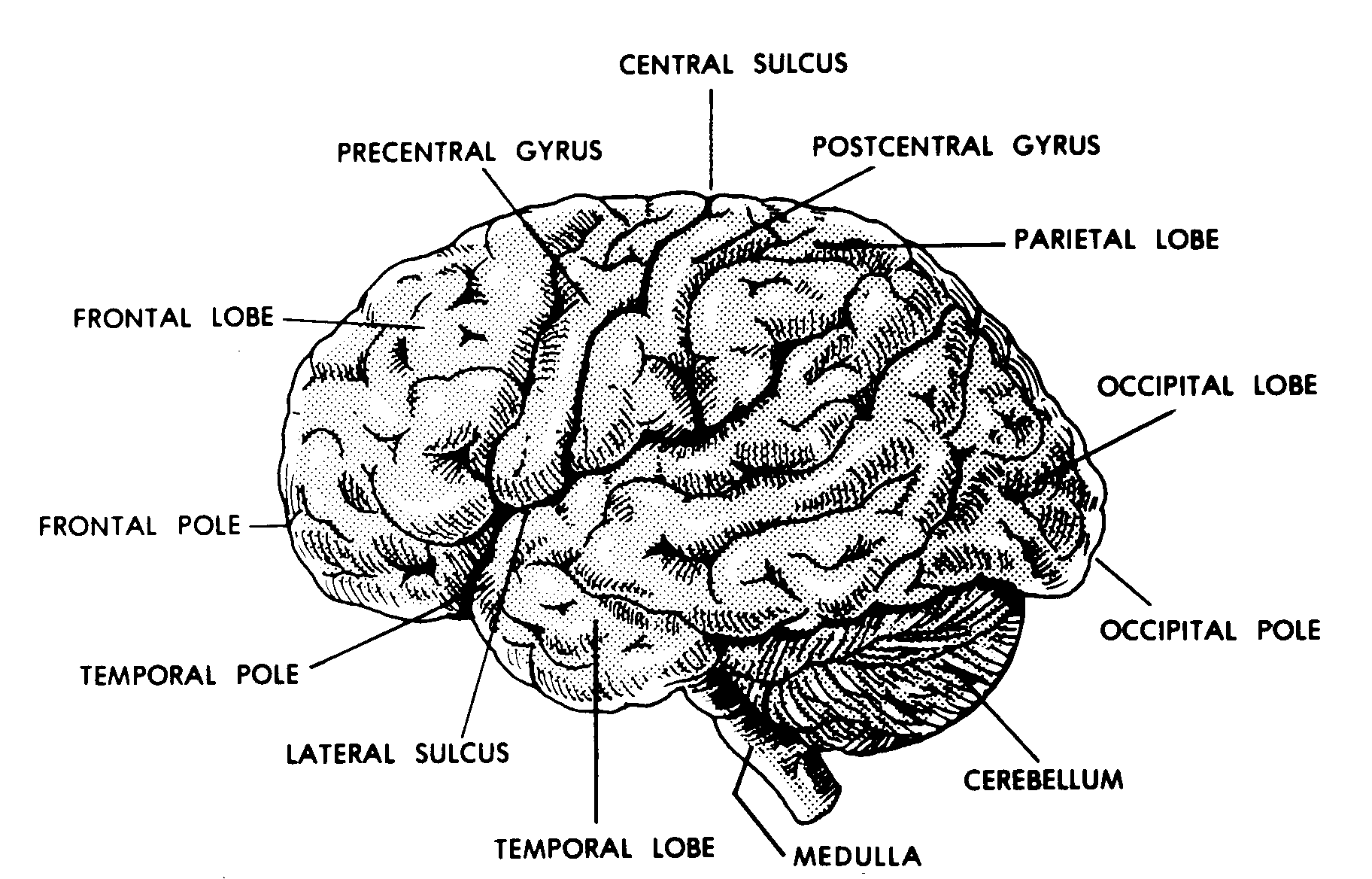
Figure 12-3. Human brain; sideview.
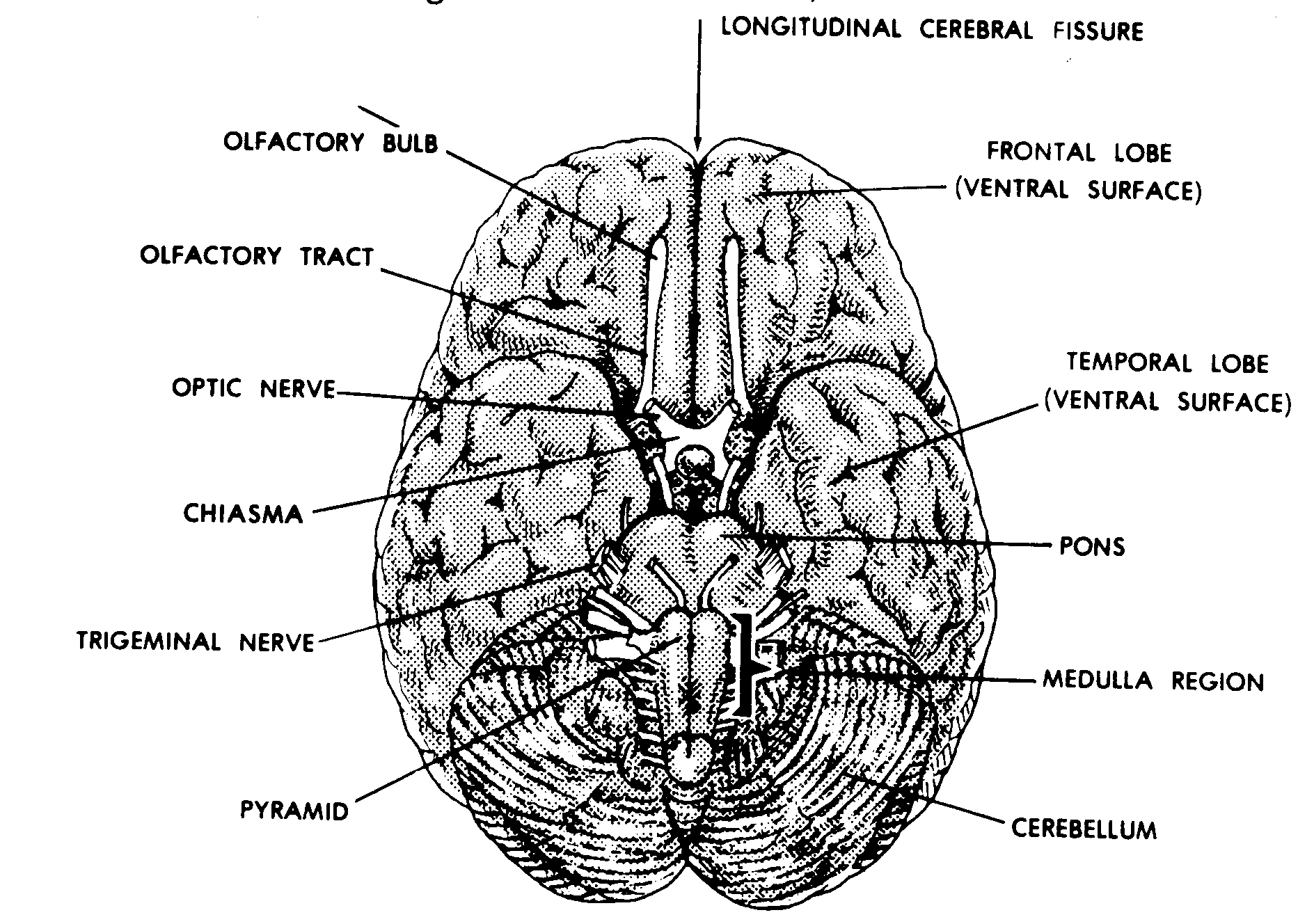
Figure 12-4. Human brain; bottom view.
c. The Cerebrum. Attached to the forebrainstem are the two cerebral hemispheres (Figure 12-5). Together, these two hemispheres make up the cerebrum. Among related species, the cerebrum is the newest development of the brain.
(1) Cerebral hemispheres. The cerebrum consists of two cerebral hemispheres, right and left. They are joined together by a very large fiber tract known as the corpus callosum (the great commissure).
(2) Lobes. Each hemisphere can be divided into four lobes. Each lobe is named after the cranial bone it lies beneath--parietal, frontal, occipital, and temporal. (Actually, there are five lobes. The fifth is hidden at the bottom of the lateral fissure. It is known as the insula or insular lobe. It is devoted mainly to visceral activities.)
(3) Gyri and sulci. The cerebral cortex, the thin layer at the surface of each hemisphere, is folded. This helps to increase the amount of area available to neurons. Each fold is called a gyrus. Each groove between two gyri is called a sulcus.
(a) The lateral sulcus is a cleft separating the frontal and parietal lobes from the temporal and occipital lobes. Therefore, the lateral sulcus runs along the lateral surface of each hemisphere.
(b) The central sulcus is a cleft separating the frontal from the parietal lobe. Roughly, each central sulcus runs from the left or right side of the cerebrum to top center and over into the medial side of the cerebrum.
(c) There are two gyri that run parallel to the central sulcus. Anterior to the central sulcus is the precentral gyrus. Posterior to the central sulcus is the postcentral gyrus.
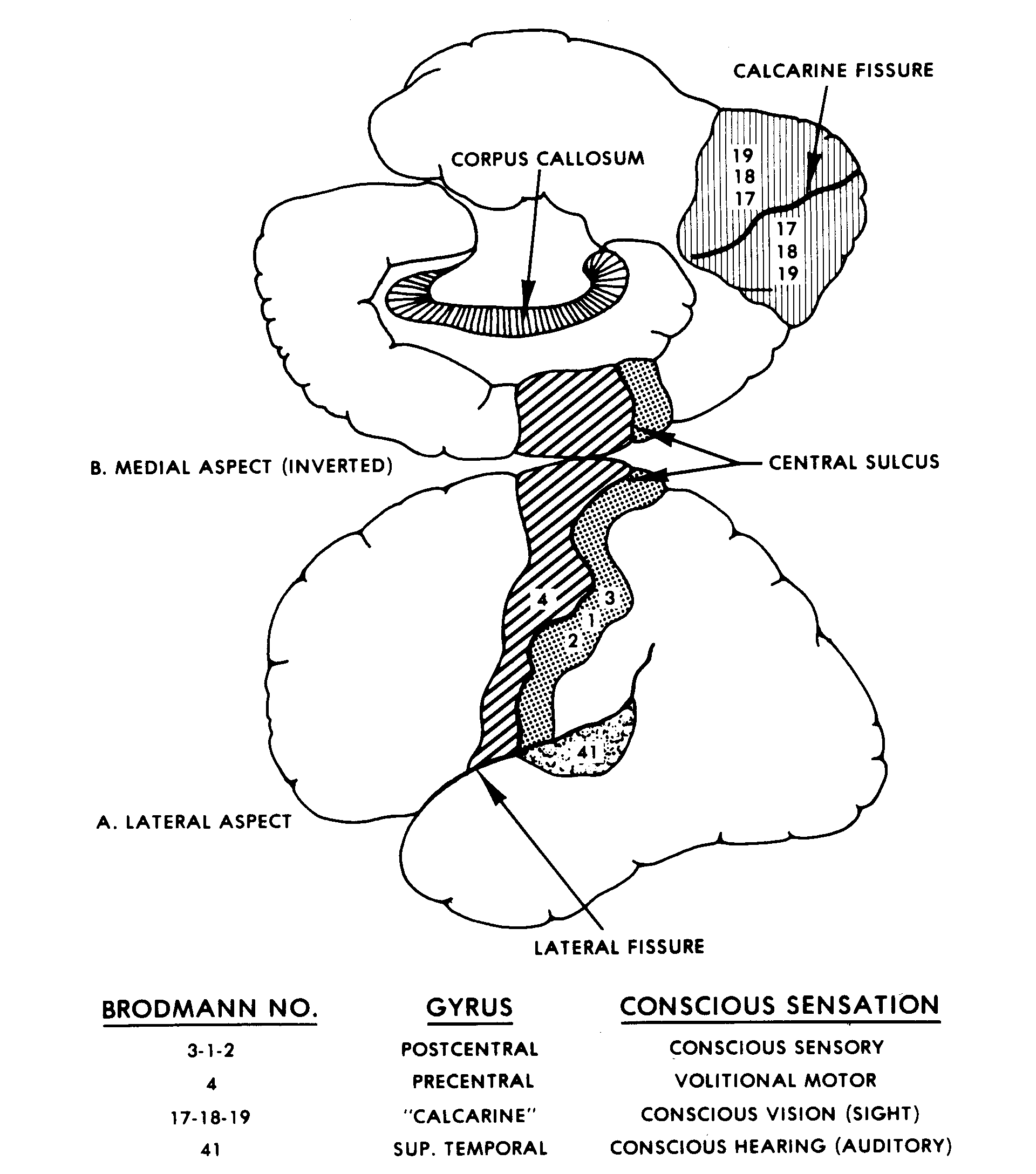
Figure 12-5. Left cerebral hemisphere.
12-7. THE HUMAN SPINAL CORD
Extending inferiorly from the brain is the spinal cord (Figure 12-6).

Figure 12-6. A cross section of the spinal cord.
a. The spinal cord is continuous with the brainstem. Together, the spinal cord and the brainstem are called the neuraxis. The foramen magnum is taken as the point that divides the brainstem from the spinal cord. Thus, the brainstem is within the cranial cavity of the skull, and the spinal cord is within the vertebral (spinal) canal of the vertebral column.
b. The spinal cord has a central portion known as the gray matter. The gray matter is surrounded by the white matter.
(1) The gray matter is made up of the cell bodies of many different kinds of neurons.
(2) The white matter is made up of the processes of neurons. The white color is due to their myelin sheaths. These processes serve several purposes: Many make a variety of connections within the spinal cord. Many ascend the neuraxis to carry information to the brain. Many descend the neuraxis to carry commands from the brain.
Section III. THE PERIPHERAL NERVOUS SYSTEM (PNS)
12-8. PERIPHERAL NERVES
Connecting the CNS to all parts of the body are individual organs known as nerves. A nerve is a collection of neuron processes together and outside of the CNS. Peripheral nerves are nerves which pass from the CNS to the periphery of the body. Together, they are referred to as the peripheral nervous system.
a. These nerves are bilateral and segmental.
(1) Bilateral. This means that the peripheral nerves occur in pairs. In each pair, there is one nerve to the right and one to the left.
(2) Segmental. The pairs of peripheral nerves occur in intervals, corresponding to the segments of the human embryo.
b. Peripheral nerves connected to the brainstem are called cranial nerves. They are numbered from I through XII and also have individual names.
c. Peripheral nerves connected to the spinal cord are called spinal nerves. They are identified by a letter representing the region of the vertebral column and a number representing the sequence in the region:
(1) Cervical: C-1 through C-8.
(2) Thoracic: T-1 through T-12.
(3) Lumbar: L-1 through L-5.
(4) Sacral: S-1 through S-5.
(5) Coccygeal.
Thus, there are 31 pairs of spinal nerves.
12-9. A "TYPICAL" SPINAL NERVE (FIGURE 12-7)
In the human body, every spinal nerve has essentially the same construction and components. By learning the anatomy of one spinal nerve, you can understand the anatomy of all spinal nerves. Like a tree, a typical spinal nerve has roots, a trunk, and branches (rami).

Figure 12-7. A typical spinal nerve, with a cross section of the spinal cord.
a. Coming off of the posterior and anterior sides of the spinal cord are the posterior (dorsal) and anterior (ventral) roots of the spinal nerve. An enlargement on the posterior root is the posterior root ganglion. A ganglion is a collection of neuron cell bodies, together, outside the CNS.
b. Laterally, the posterior and anterior roots of the spinal nerve join to form the spinal nerve trunk. The spinal nerve trunk of each spinal nerve is located in the appropriate intervertebral foramen of the vertebral column. (An intervertebral foramen is a passage formed on either side of the junction between two vertebrae.)
c. Where the spinal nerve trunk emerges laterally from the intervertebral foramen, the trunk divides into two major branches. These branches are called the anterior (ventral) and posterior (dorsal) primary rami (ramus, singular). The posterior primary rami go to the back. The anterior primary rami go to the sides and front of the body, and to the upper and lower members.
Section IV. THE AUTONOMIC NERVOUS SYSTEM
12-10. CONTROL OF VISCERAL ACTIVITIES
The autonomic nervous system (ANS) is that portion of the nervous system concerned with commands for smooth muscle tissue, cardiac muscle tissue, and glands.
a. The term visceral organs may be used to include:
(1) The various hollow organs of the body whose walls have smooth muscle tissue in them. Examples are the blood vessels and the gut.
(2) The glands.
b. The visceral organs are innervated by the ANS. This results in a "visceral motor system." For most of us, the control of the visceral organs is automatic, that is, without conscious control. However, recent research demonstrates that conscious control of some of the visceral organs is possible after proper training.
12-11. TWO MAJOR SUBDIVISIONS
The ANS is organized into two major subdivisions--the sympathetic and the parasympathetic nervous systems.
a. The neurons of the sympathetic nervous system originate in the thoracic and lumbar regions of the spinal cord. Thus, it is also known as the thoraco-lumbar outflow.
b. Some of the neurons of the parasympathetic nervous system originate in nuclei of the brainstem. Others originate in the sacral region of the spinal cord. Thus, the parasympathetic nervous system is also known as the cranio-sacral outflow.
c. In the ANS, there are always two neurons (one after the other) connecting the CNS with the visceral organ. The cell bodies of the second neurons form a collection outside the CNS, called a ganglion. Processes of these postganglionic neurons extend to the visceral organs. Those processes going to peripheral visceral organs are included with the peripheral nerves.
12-12. EQUILIBRIUM
Under ordinary circumstances, the sympathetic and parasympathetic nervous system have opposite effects upon any given visceral organ. That is, one system will stimulate the organ to action, and the other system will inhibit it. The interplay of these two systems helps visceral organs to function within a stable equilibrium. This tendency to produce an equilibrium is called homeostasis.
12-13. RESPONSE TO STRESS
Under conditions of stress, the sympathetic nervous system produces a "fight-or-flight" response. In other words, it mobilizes all of the energy producing structures of the body. Simultaneously, it inhibits those structures that do not contribute to the mobilization of energy. For example, the sympathetic nervous system makes the heart beat faster. Later, as equilibrium is restored, the parasympathetic nervous system slows the heart down.
Section V. ELECTROCHEMICAL TRANSMISSION OF NEURON IMPULSES
2-14. INTRODUCTION
a. The functional elements of the human nervous system are the neurons. The neurons are alined in sequences, one neuron after the other, to form circuits. The transmission of information along the length of a neuron is electrochemical in nature.
b. An important fact is that "connecting" neurons do not actually touch each other. Instead, there is a space between the end of one and the beginning of the next ("continuity without contact"). A specified chemical, called a neurotransmitter, is required to cross the gap between one neuron and the next.
12-15. RESTING POTENTIAL
As a part of their life processes, neurons are able to produce a concentration of negative ions inside and a concentration of positive ions outside of the cell membrane. The difference in the concentration of ions produces an electrical potential across the membrane. This condition is often referred to as polarization. When the neuron is not actually transmitting, this electrical potential across the membrane is known as the resting potential.
12-16. ACTION POTENTIAL (DEPOLARIZATION AND REPOLARIZATION)
Where a stimulus is applied to the neuron, the polarity of the ions is disrupted at the same location. Thus, that location is said to be depolarized. The ions in adjacent areas along the neuron then attempt to restore the original polarity at the location of the stimulus. However, as repolarization occurs in the area of the stimulus, the adjacent areas themselves become depolarized. This results in a wavelike progression of depolarization/repolarization along the length of the neuron. By this means, information is transferred along the neuron.
12-17. EFFECT OF THE THICKNESS OF THE NEURON PROCESSES
The speed with which an impulse travels is proportional to the thickness of the neuron process. The thickest processes (A fibers) have the fastest transmission (about 120 meters/second). The thinnest processes (C fibers) are the slowest (as slow as 1/2 meter/second). The B fibers (thicker than C fibers and thinner than A fibers) are faster than C fibers and slower than A fibers.
12-18. THE SYNAPSE
The gap between successive neurons is wide enough that impulses do not travel from one neuron to the next in the same way as along a single neuron. Information travels from one neuron to the next by means of a chemical neurotransmitter. Together, the gap and the "connecting" membranes of the neurons are called the synapse (Figure 12-8). The gap is called the synaptic cleft.
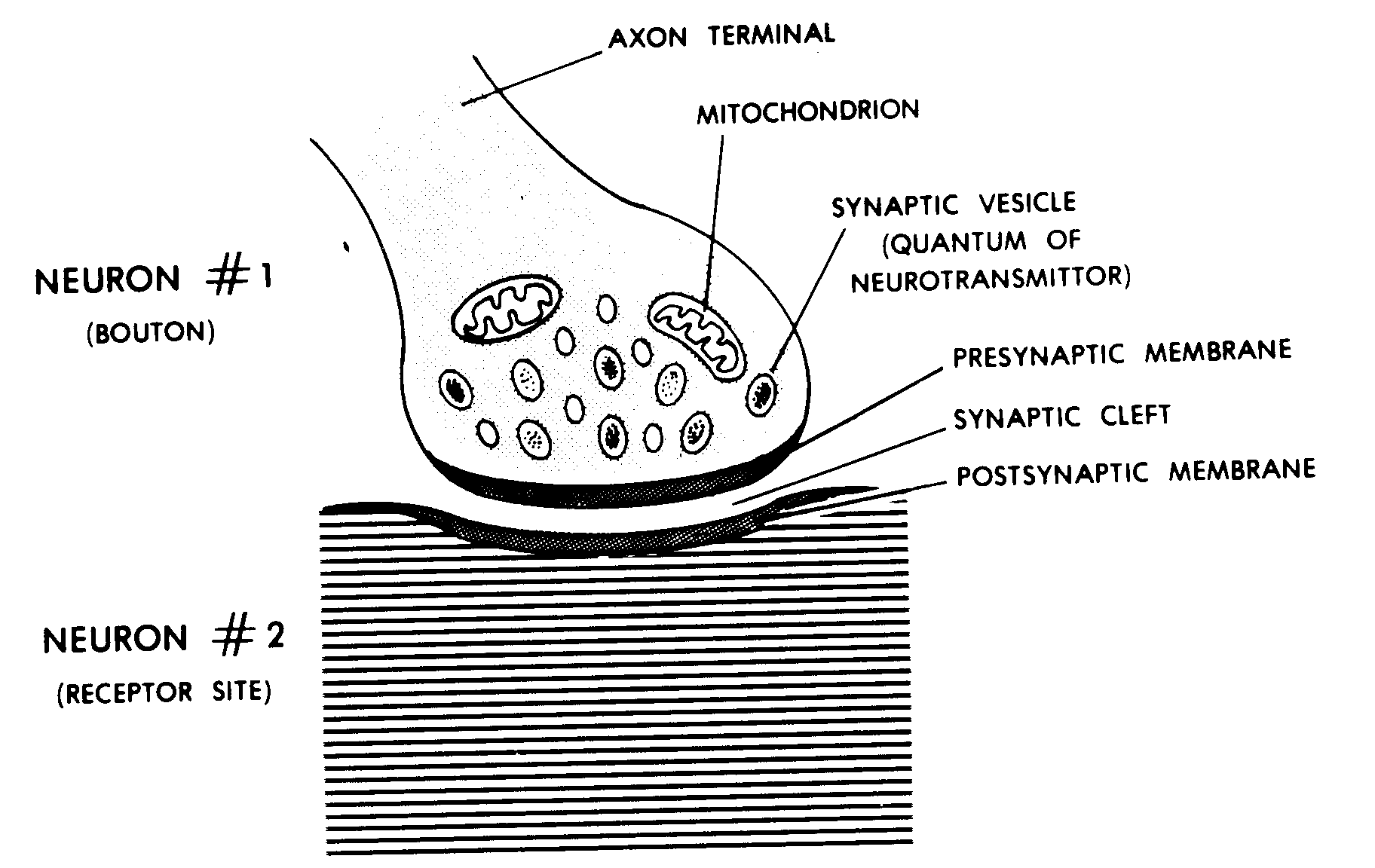
Figure 12-8. A synapse.
a. Many synaptic vesicles (bundles of neurotransmitters) are found in the terminal bulb (bouton) of the first neuron. Each vesicle contains a quantum, a specific amount, of neurotransmitter or a substance used to make the neurotransmitter.
b. When the impulse reaches the bouton, these vesicles are stimulated to release their neurotransmitter. The neurotransmitter then passes out of the bouton, through the presynaptic membrane, into the synaptic cleft. On the other side of the synaptic cleft is the postsynaptic membrane. This is the receptor site of the second neuron.
c. The neurotransmitter is located only in the terminal bulb of the first neuron. For this reason, impulses travel in only one direction through the synapse, from the first to the second neuron. Since this process consumes much energy, there are many well-developed mitochondria in the bouton, or terminal bulb.
12-19. THE NEUROMUSCULAR JUNCTION
While the synapse is the "connection" between two neurons, the neuromuscular junction (Figure 12-9) is the "connection" between a motor neuron and a striated muscle fiber.
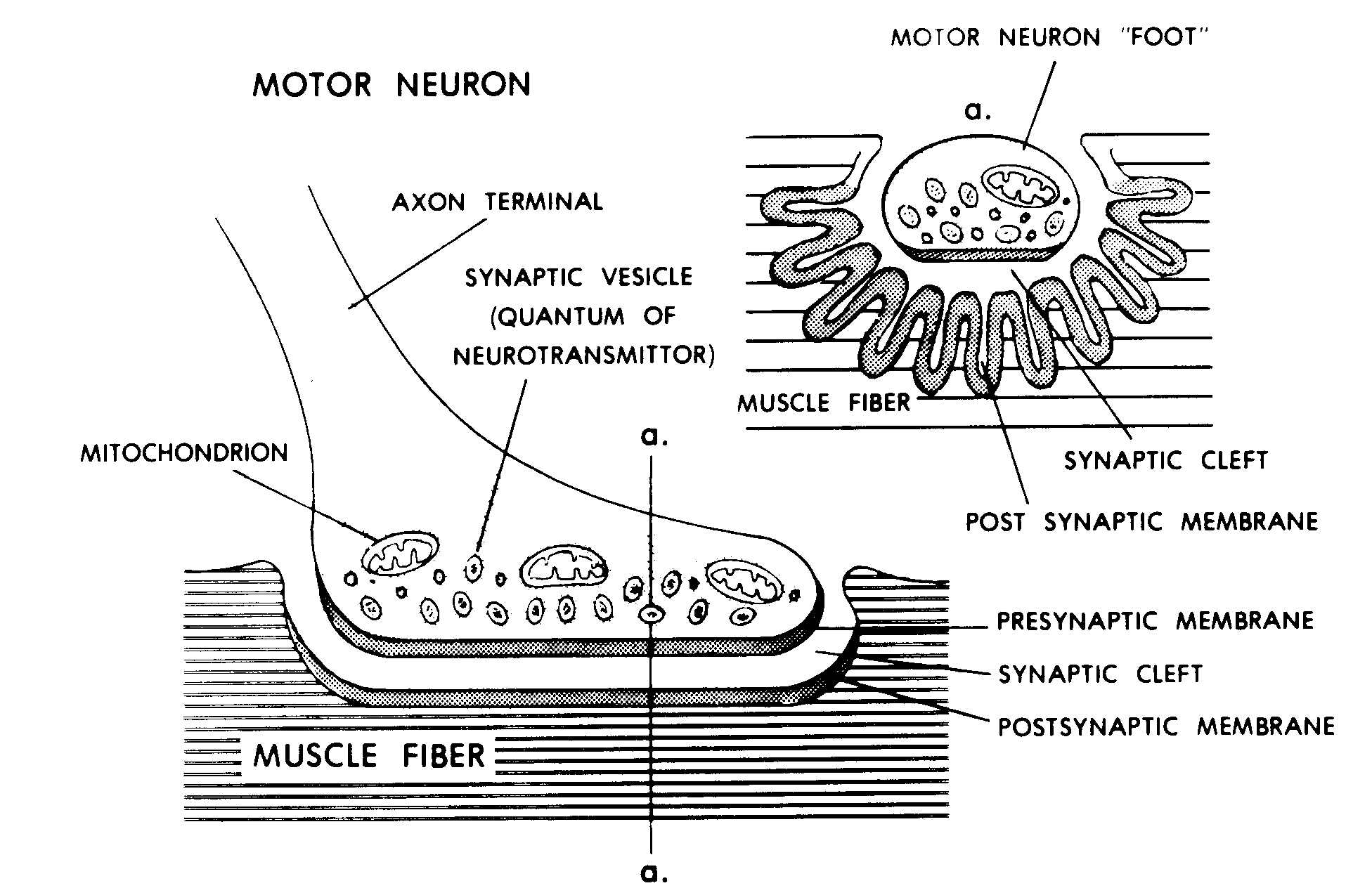
Figure 12-9. A neuromuscular junction.
a. In general terms, the neuromuscular junction and the synapse are physiologically identical. Synaptic vesicles in the enlarged bouton of the motor neuron contain the neurotransmitter acetylcholine (ACH). As an impulse reaches the bouton, ACH is released and passes through thepresynaptic membrane into the synaptic cleft. However, the surface of the postsynaptic membrane is in a series of longitudinal folds. This greatly increases the surface area receptive to the ACH.
b. The motor unit is the group of striated muscle fibers innervated by the terminal arborization (tree-like branching) of one motor neuron. The fewer the muscle fibers found per motor unit, the more the muscle is capable of finer movements. As the number in the motor unit increases, the muscle action is coarser. When a muscle is to be used, the nervous system recruits just enough motor units to supply the strength needed for the work to be done.
Section VI. THE GENERAL REFLEX AND THE REFLEX ARC
12-20. THE GENERAL REFLEX
The simplest reaction of the human nervous system is the reflex. A reflex is defined as an automatic reaction to a stimulus.
12-21. THE GENERAL REFLEX ARC
The pathway followed by the stimulus (impulse) from beginning to end is the reflex arc. The general reflex arc (Figure 12-10) of the human nervous system has a minimum of five components:
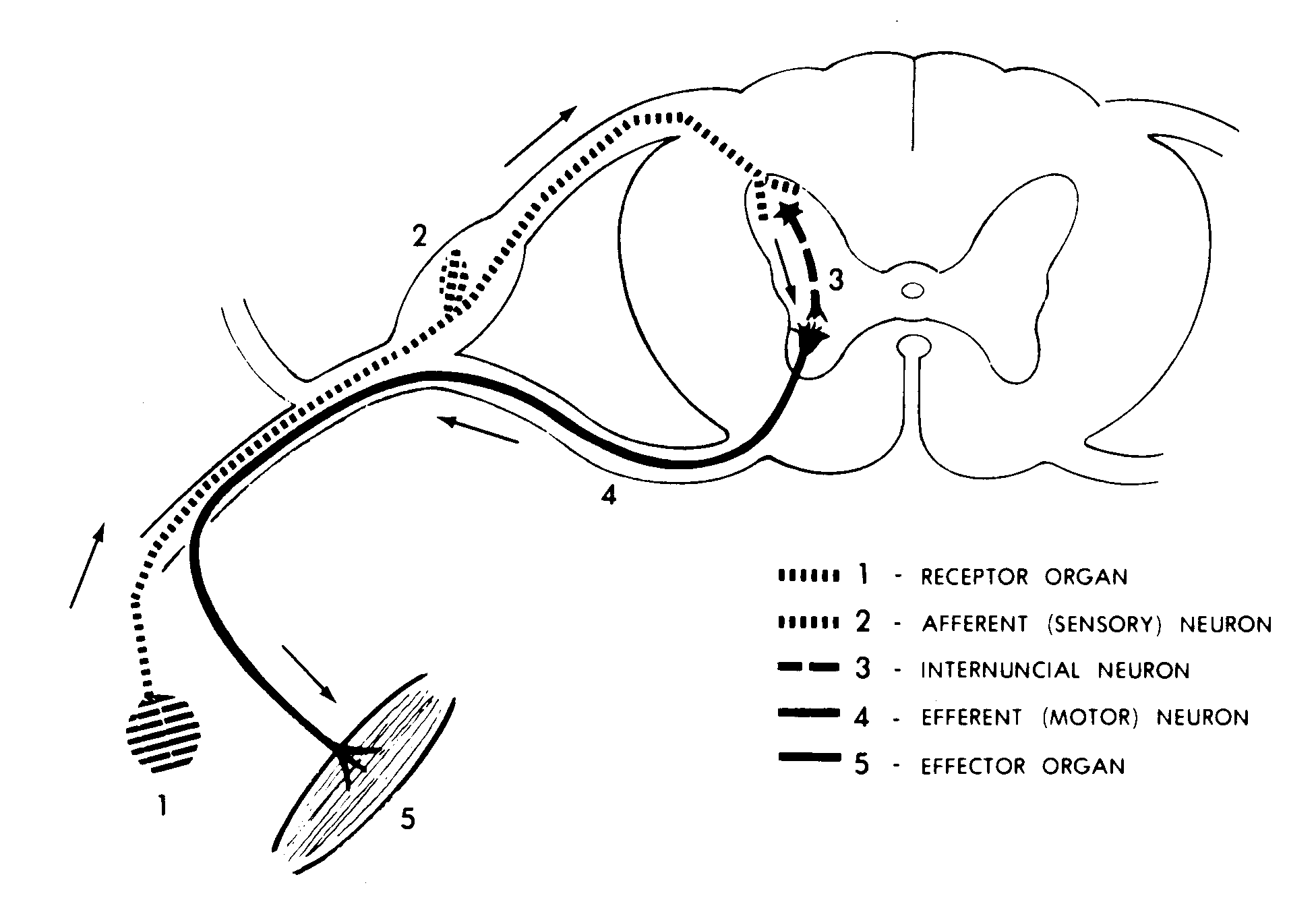
Figure 12-10. The general reflex arc.
a. The stimulus is received by a receptor organ specific to that stimulus.
b. From the receptor organ, the stimulus is carried to the CNS by way of an afferent (sensory) neuron within the appropriate peripheral nerve. The cell body of this afferent neuron is located in the posterior root ganglion of a spinal nerve or the individual ganglion of a cranial nerve.
c. Within the spinal cord or brainstem, the terminal of the afferent neuron synapses with the interneuron, or internuncial neuron.
INTER = between
NUNCIA = messenger
In turn, the internuncial neuron synapses with the cell body of the efferent (motor) neuron.
d. In the spinal cord, the cell bodies of the efferent (motor) neurons make up the anterior column of the gray matter. In the brainstem, the motor neurons make up the individual nuclei of the cranial nerves. The axon of the motor neuron passes out of the CNS by way of the appropriate peripheral nerve. Command information is thus carried away from the CNS.
e. The information is then delivered by the motor neuron to the effector organ. Somatic motor neurons lead to striated muscle fibers, particularly in skeletal muscles. Autonomic (visceral) motor neurons lead to smooth muscle tissue, cardiac muscle tissue, or glands.
Section VII. GENERAL SENSORY PATHWAYS OF THE HUMAN NERVOUS SYSTEM
12-22. INTRODUCTION TO PATHWAYS
A pathway of the human nervous system is the series of neurons or other structures used to transmit an item of information. In general, we consider two major types of pathways--the general sensory pathways and the motor pathways.
a. Ascent or Descent Through the Neuraxis. The general sensory pathways ascend through the neuraxis to the brain. The motor pathways descend through the neuraxis from the brain. The neuraxis includes both the spinal cord and the brainstem. The pathways are included in various fiber tracts of the neuraxis.
b. Crossing to the Opposite Side (Decussation). At some specific level in the neuraxis, all of these pathways cross to the opposite side of the midline of the CNS. (Each crossing is called a decussation.) Thus, the right cerebral hemisphere of the brain communicates with the left half of the body. The left cerebral hemisphere communicates with the right half of the body.
12-23. INTRODUCTION TO GENERAL SENSORY PATHWAYS
a. The General Senses. The general senses detect those specific stimuli which are received throughout the body (general distribution). When these general senses are perceived at the conscious level (in the cerebral cortex), they are known as sensations. The general senses of humans include pain, touch, temperature, and proprioception ("body sense").
b. Neurons of a General Sensory Pathway. A general sensory pathway extends from the point where the stimulus is received to the postcentral gyrus of the cerebral hemisphere (para 12-6c(3)(c)). The postcentral gyrus is the site of conscious sensation of a stimulus. Between the point of stimulus reception and the postcentral gyrus, there is a minimum of three neurons in series.
(1) The first neuron is the afferent (sensory) neuron. It picks up the information from the sensory receptor organ and carries it to the CNS via the appropriate peripheral nerves.
(2) The second neuron is the interneuron, located within the spinal cord or brainstem. It crosses the midline of the CNS to the opposite side. It then ascends the neuraxis to the forebrainstem, where it reaches a mass of gray matter called the thalamus. In the thalamus, the interneuron synapses with the cell body of the third neuron.
(3) The axon of the third neuron projects up through the cerebral hemisphere to the appropriate location in the postcentral gyrus.
c. Homunculus of Conscious Sensations. There is a specific location in the postcentral gyrus which corresponds to each location in the body. For example, a location in the postcentral gyrus near the midline of the brain (at the top of the cerebral hemisphere) receives information from the hip region. On the other hand, information from the tongue and the pharynx projects to the lowest part of the postcentral gyrus, just above the lateral sulcus.
d. Visceral Sensory Inputs. Visceral sensory inputs follow pathways different from those of other general sensory pathways. The inputs for visceral reflex actions usually travel via the parasympathetic nerves. The visceral inputs for pain usually travel via the sympathetic nerves.
12-24. PAIN--A GENERAL SENSE
Pain is an ancient protective mechanism which generally helps us to avoid injury. However, tolerance for pain varies from one individual to another.
a. Means of Reducing Pain (Analgesia).
(1) Endorphins ("morphine from within"). Endorphins are chemicals found naturally within the body which tend to block the sensation of pain.
(2) Drugs. Clinically, a number of drugs are used to block or reduce the sensation of pain.
(3) Competing inputs. Competing pain stimuli tend to minimize each other. The body usually recognizes one pain stimulus at a time. Thus, an individual may "bite his lip" when he anticipates a painful experience.
b. Pain Receptor. The pain receptor is not a specific receptor organ, as with most senses. This receptor is referred to as a free nerve ending.
c. Excessive Stimulation. If any of the other senses receives excessive stimulus, pain results. Examples are excessive light and excessive noise.
d. Pain Reflex Arc. Generally, a pain sensory input causes a reflex action long before the information reaches the cerebral cortex and the pain is consciously perceived. For example, you will remove your hand from a hot object before you realize you have been burned.
e. Pathway for Conscious Sensation of Pain. As usual, the pathway leading to conscious sensation of pain consists of three neurons.
(1) The first neuron is the afferent (sensory) neuron from the free nerve ending. Within the CNS, it synapses with the interneuron.
(2) The axon of the interneuron crosses to the opposite side of the CNS. It then ascends the neuraxis in a fiber tract known as the lateral spinothalamic tract. This tract is found in the lateral funiculus (see Figure 12-6). In the thalamus, the interneuron synapses with the third neuron.
(3) The third neuron projects to the appropriate location of the postcentral gyrus of the cerebral hemisphere. Here, this information is interpreted or recognized as a pain sensation from a particular part of the body.
12-25. TEMPERATURE -- GENERAL SENSES
There are two categories of temperature in the body--warmth and cold.
a. However, these are relative entities. For example, a given temperature seems cool when compared to a much higher temperature and seems hot when compared to a much lower temperature.
b. In addition, the body has two different mechanisms for sensing temperature.
(1) Specific sensory receptors detect warmth and especially cold in the periphery of the body.
(2) Special heat-sensitive neurons in the hypothalamus detect increases in the temperature of the blood that flows through the hypothalamus (portion of the forebrainstem). By this means, the body monitors the core temperature, the temperature in the central part of the body.
c. Neurons for the general sense of temperature use pathways similar to those discussed for pain (para 12-24e). They include both nerves and fiber tracts.
12-26. TOUCH -- GENERAL SENSES
Throughout the body are a variety of sensory receptors which detect varying degrees of pressure. For example, the pacinian corpuscles are typical of the receptors which detect deep pressure. In addition, an individual can usually identify the location of a touch on his body; in fact, he can usually distinguish two simultaneous touches to adjacent areas (the "two-touch test"). As usual with the general senses, sensory inputs for touch can also result in immediate reflex actions.
a. Pathway for Conscious Sensation of Light Touch.
(1) The pathway for the conscious sensation of light touch begins with the usual afferent (sensory) neuron as the first neuron. The afferent neuron carries the information to the CNS by way of the appropriate nerve.
(2) In the CNS, the afferent neuron synapses with the interneuron, the second neuron of the pathway. After crossing to the opposite side of the CNS, the interneuron ascends the neuraxis in the fiber tract known as the anterior spinothalamic tract. This is in the anterior funiculus of the spinal cord (Figure 12-6).
(3) In the thalamus, the second neuron synapses with the third neuron. The axon of the third neuron then projects to the appropriate location in the postcentral gyrus of the cerebral hemisphere. There, it is interpreted as the conscious sensation, light touch.
b. Pathway for Conscious Sensation of Deep Touch. The pathway for deep touch is quite different from that for light touch.
(1) Still, the first neuron is the afferent neuron from the deep touch receptor to the CNS via the appropriate nerve. When the axon of the afferent neuron enters the CNS, it turns upward and ascends the neuraxis in the posterior funiculus (Figure 12-6) of the same side that it entered. In other words, it does not yet cross the midline of the CNS.
(2) In the lower hindbrainstem, the axon of the first neuron synapses with the cell body of the second neuron. The axon of the second neuron then crosses to the opposite side of the brainstem. This axon then continues the ascent through the neuraxis to the thalamus, where it synapses with the third neuron.
(3) Again, the axon of the third neuron projects to the appropriate location in the postcentral gyrus of the cerebral hemisphere. There, impulses are interpreted as conscious sensations of deep touch.
12-27. "BODY SENSE"
a. General. Body sense is the combined information from a number of sensory inputs. Second by second, these inputs keep the brain informed of the specific posture of the body and its parts. Some of the senses involved include:
(1) Muscle sense (proprioception).
(2) Joint capsule sense.
(3) Integument senses.
(4) Special senses (eye, ear, etc.).
b. Proprioception (Muscle Sense).
(1) For proprioception, there is a very special receptor organ to monitor the degree of stretch of the muscle. These receptor organs, called muscle spindles or stretch receptors, are distributed within the fleshy belly of each skeletal muscle. In effect, the muscle spindles are parallel to striated muscle fibers of the skeletal muscles. Therefore, as the muscle fibers contract or are stretched, the muscle spindle detects relative muscle length.
(a) The afferent neuron from the muscle spindle is known as the annulospiral neuron because its terminal is coiled. Due to this coiling, it is a spring-like apparatus which can be stretched or compressed according to the condition of the muscle. The annulospiral neuron travels to the CNS by way of the appropriate nerve. It continuously carries information about the specific state of the muscle.
(b) An annulospiral neuron from a muscle in one of the limbs, in particular, synapses directly on the motor neuron that carries commands back to the same muscle. This motor neuron is called the alpha motor neuron. Together, the annulospiral neuron and the alpha motor neuron make up the stretch (monosynaptic) reflex. Due to this reflex, there is a proportionate increase in the tension of a muscle as it stretches.
(2) Another stretch receptor associated with the skeletal muscle is the Golgi tendon organ. As its name implies, this organ is located within the tendon of the muscle. The Golgi tendon organ is located in the tendon near its attachment to the muscle fibers. Thus, it detects relative muscle tension. Its threshold is higher than that of the muscle spindles; in other words, there must be proportionately more contraction before it puts out a signal. Thus, when the muscle has been stretched excessively and might be subject to injury, its afferent neuron carries the message to the CNS. This results in relaxation of the muscle.
(3) The pathway for the conscious sensation of these stretches uses the same structures as the deep touch general sense.
Section VIII. MOTOR PATHWAYS IN THE HUMAN NERVOUS SYSTEM
12-28. INTRODUCTION
The CNS receives information through the sensory pathways and collates this information against information stored in memory. This results in a decision. If the decision is to do something, then the CNS sends out commands through the motor pathways to the effector organs (muscles, glands, etc.).
a. The motor pathways descend in the neuraxis and transmit the commands to the motor neurons. The processes of motor neurons leave the CNS by way of the peripheral nerves. The somatic motor neurons activate striated muscle fibers. The visceral motor neurons activate smooth muscle tissue, cardiac muscle tissue, and glands.
b. We usually consider two general motor pathways--the pyramidal motor pathways and the extrapyramidal motor pathways.
12-29. PYRAMIDAL MOTOR PATHWAYS
A pyramidal motor pathway is primarily concerned with volitional (voluntary) control of the body parts, particularly with the fine movements of the hands. Since such a pathway is concerned with volitional actions, it is suitable for neurological screening and testing.
a. Cerebral Motor Cortex. The pyramidal motor pathway begins in the precentral gyrus of the cerebral hemisphere. As we have already seen with the sensory pathways, the neurons making up the cerebral cortex of the precentral and postcentral gyri are arranged in a pattern (motor homunculus) corresponding to the various parts of the body to which they are connected.
b. Motor Neurons. From the precentral gyrus, the axons of these upper motor neurons (UMN) pass into the neuraxis of the CNS and descend. At the level of the appropriate segmental nerve, the UMN synapses either directly or indirectly with a lower motor neuron of the segmental nerve. Direct synapses (monosynaptic) provide the most rapid reactions. Such direct synapses are used in particular for the fine movements of the hands.
c. Corticospinal Pathways. The medulla is the lowest part of the brainstem. On the underside of the medulla, the axons of the UMNs form a pair of structures known as the pyramids. Immediately below the pyramids, at the beginning of the spinal cord, the axons cross to the opposite side of the CNS (spinal cord). The axons then descend as the lateral corticospinal tract, within the lateral funiculus (Figure 12-6). Thus, the left cerebral hemisphere commands the right side of the body, and the right cerebral hemisphere controls the left side of the body.
12-30. EXTRAPYRAMIDAL MOTOR PATHWAYS
The extrapyramidal motor pathways are concerned with automatic (nonvolitional) control of body parts. This particularly includes patterned, sequential movements or actions. Thus, the major command system of the human nervous system uses these pathways. There are several extrapyramidal motor pathways. Having multisynaptic circuits throughout the CNS, they use many intermediate relays before reaching the effector organs. The cerebellum of the brain plays a major role in extrapyramidal pathways; the cerebellum is the major center for coordinating the patterned sequential actions of the body, such as walking.
Section IX. LEVELS OF CONTROL IN THE HUMAN NERVOUS SYSTEM
12-31. INTRODUCTION
a. General Concept. The human nervous system can be thought of as a series of steps or levels (Figure 12-11). Each level is more complex than the level just below. No level is completely overpowered by upper levels, but each level is controlled or guided by the next upper level as it functions.
b. Changes With Development or Injury.
(1) Babinski's reflex involves dorsiflexion of the big toe when the sole of the foot is stimulated. It can be normally observed in infants up to 18 months of age. As the pyramidal motor pathways develops completely, this reflex disappears. However, if the pyramidal motor system is injured, the Babinski reflex tends to return.
(2) Thus, it is possible to evaluate the extent of development of an individual by identifying the highest level of control. In the case of injury, the highest active level of control helps determine the site of the injury.
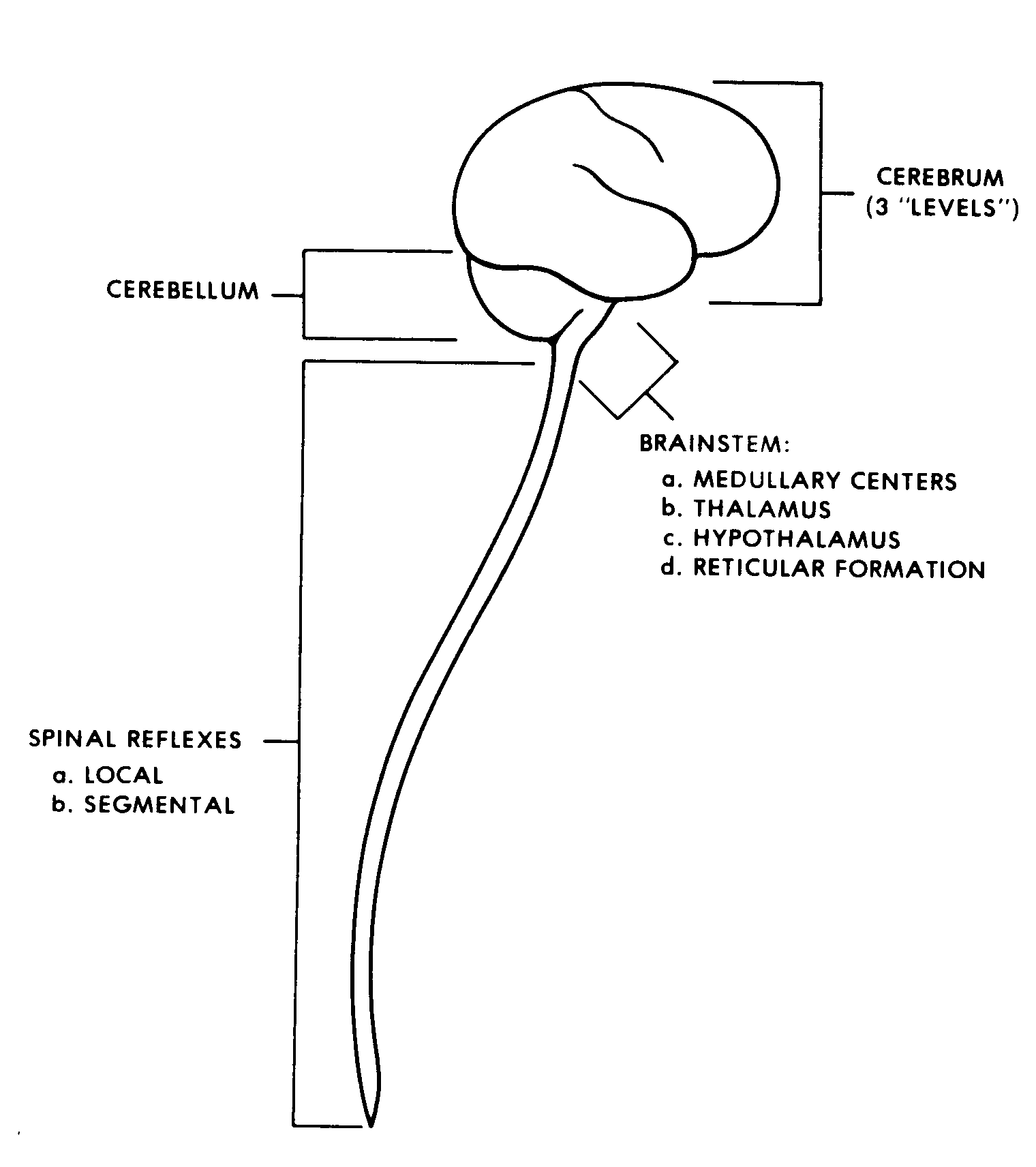
Figure 12-11. Levels of the CNS.
12-32. REFLEXES
a. Reflex Arc. The simplest and lowest level of control is the reflex arc. The reflex arc operates essentially on the level of the sensory input.
b. Segmental Reflexes. Segmental reflexes produce a wider reaction to a stimulus than the reflex arc. For this purpose, the nervous system is organized more complexly. Thus, information spreads to a wider area of CNS. We can observe a greater reaction to the stimulus.
12-33. BRAINSTEM "CENTERS"
Within the brainstem, there is a well integrated series of control centers.
a. Visceral Centers of the Medulla. There is a group of nuclei in the medulla of the hindbrainstem. Together, these nuclei control the visceral activities of the body, such as respiration, heart beat, etc.
b. Reticular Formation. Within the substance of the brainstem is a diffuse system called the reticular formation. The reticular formation has a facilitory (excitatory) area and an inhibitory area. Thus, this control area tends to activate or slow down activities of the body. Thus, it is responsible for producing sleep or wakefulness.
c. Hypothalamus and Thalamus.
(1) The thalamus is a group of nuclei found together in the forebrainstem. The thalamus is the major relay center of sensory inputs from the body.
(2) The hypothalamus is a higher control center for visceral activities of the body. It is found associated with the thalamus.
12-34. CEREBELLUM
The cerebellum has been greatly developed, with many functional subdivisions. It is the primary center for the integration and control of patterned, sequential motions of the body. The cerebellum is also the center of control of body posture and equilibrium.
12-35. CEREBRUM
In humans, the highest level of nervous control is localized in the cerebrum. It is at this level that conscious sensation and volitional motor activity are localized. Even so, we can clearly designate three levels of control within the cerebrum:
a. Visceral (Vegetative) Level. This level is concerned primarily with visceral activities of the body, as related to fight-or-flight, fear, and other emotions.
b. Patterned (Stereotyped) Motor Actions. Here, activities of the body are standardized and repetitive in nature. An example of a stereotyped pattern of muscle activity would be the sequence of muscle actions involved in walking.
c. Volitional Level. The volitional level is the highest and newest level of control. Here, cognition (thinking) occurs, and unique, brand-new solutions can be created.
Section X. MISCELLANEOUS TOPICS
12-36. CEREBRAL AREAS
Specific areas of the cerebral cortex are concerned with specific parts of the body, with specific types of inputs, and with specific types of activities. Most often, each area is numbered as a specific Brodmann's area. For example, the precentral gyrus, concerned with volitional motions, is Brodmann's area number 4. It is the beginning of the pyramidal motor system. Likewise, the superior temporal gyrus (at the inferior margin of the lateral sulcus) is Brodmann's area number 41; it is the center for hearing.
12-37. DOMINANCE
a. About 90% of humans are right-handed. Thus, for these individuals, the left cerebral hemisphere is said to be dominant over the right cerebral hemisphere.
b. For 96% of humans, the speech center is located in the left cerebral hemisphere.
c. Thus, an injury to the left cerebral hemisphere is generally more serious than an injury to the right cerebral hemisphere.
12-38. MEMORY
a. Memory is that faculty which enables an individual to store and retrieve factual items (sensations, impressions, facts, and ideas). Memory is ultimately the result of the unceasing flow of sensory information into the CNS. These items are stored in the CNS; just exactly how and where is the subject of much research and discussion. All sensory inputs are collated against these stored items in order to arrive at an appropriate action decision. (Often, no action is the most appropriate decision.)
b. At present, at least two types of memory are recognized in the human brain--short-term memory and long-term memory.
(1) Short-term memory. A common example of short-term memory is the ability to hold a phone number in mind for a number of seconds without "memorizing" it. Short-term memory is usually limited to about seven bits of information.
(2) Long-term memory. A portion of the cerebral cortex known as the hippocampus is thought to be important in transferring information from short-term memory to long-term memory. If the hippocampus is nonfunctional, the individual can learn nothing, but his previously long-term memory remains intact.