LESSON ASSIGNMENT
LESSON 13 The Special Senses.
LESSON ASSIGNMENT Paragraphs 13-1 through 13-24.
LESSON OBJECTIVES After completing this lesson, you should be able to:
13-1. Identify functions of structures related to the special senses.
13-2. Given a list of statements about the physiology of the special senses, identify the false statement.
SUGGESTION After completing the assignment, complete the exercises at the end of this lesson. These exercises will help you to achieve the lesson objectives.
LESSON 13
THE SPECIAL SENSES
Section I. INTRODUCTION
13-1. GENERAL VERSUS SPECIAL SENSES
a. The human body is continuously bombarded by all kinds of stimuli. Certain of these stimuli are received by sense organs distributed throughout the entire body. These are referred to as the general senses.
b. Certain other stimuli (table 13-1) are received by pairs of receptor organs located in the head. These are the special senses.
|
SPECIAL SENSE |
RECEPTOR ORGAN |
STIMULUS |
|
Sight (vision) |
bulbus oculi (eye) |
light rays |
|
Hearing (audition) |
ear (cochlea) |
sound waves |
|
Balance (equilibrium) |
ear (membranous labyrinth) |
gravity |
|
Smell (olfaction) |
olfactory hair cells in nose |
airborne molecules |
|
Taste (gustation) |
taste buds in mouth |
fluid-borne molecules |
Table 13-1. The special senses.
c. Since the general senses respond to immediate contact, they are very short range. In contrast, the special senses are long range.
13-2. INPUT TO BRAIN
From the special sense organs, information is sent to the brain through specific cranial nerves. When this information reaches specific areas of the cerebral cortex, the sensations are perceived at the conscious level.
Section II. THE SPECIAL SENSE OF VISION
13-3. THE RETINA
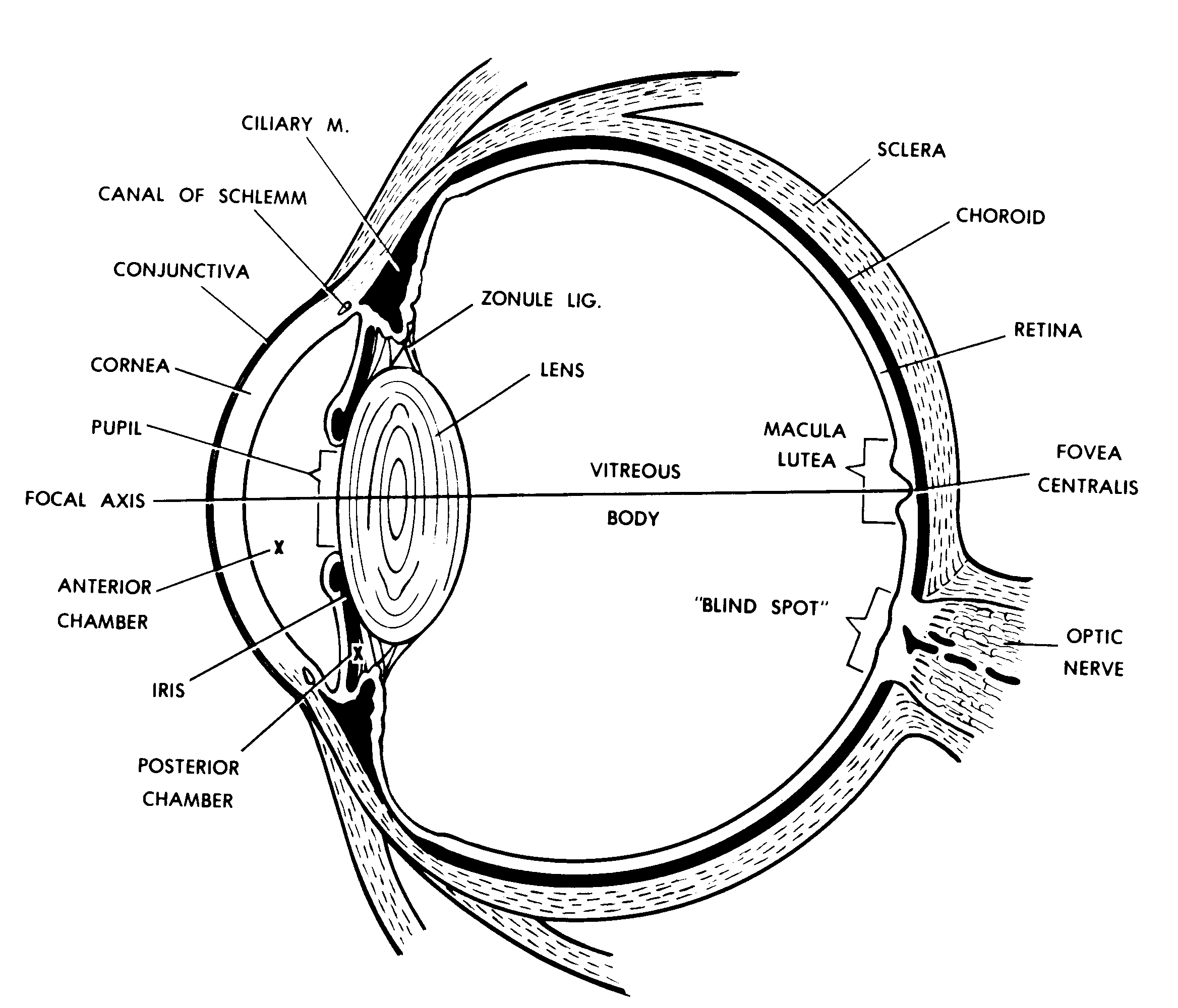
Within the bulbus oculi (eyeball) is an inner layer called the retina. See Figure 13-1 for the location of the retina within the bulbus oculi. See Figure 13-2 for the types of cells found within the retina.
Figure 13-1. A focal-axis section of the bulbus oculi.
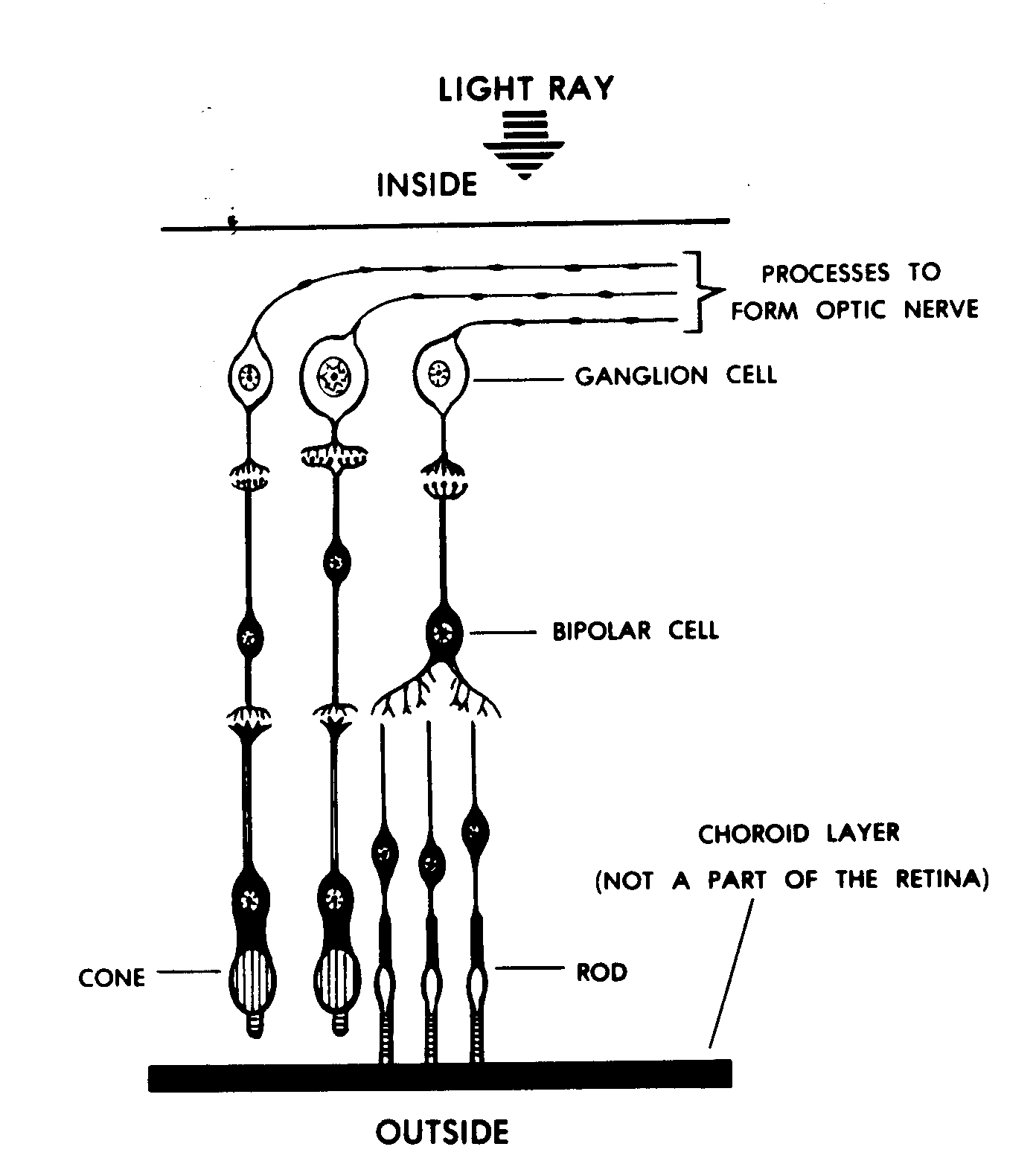
Figure 13-2. Cellular detail of the retina.
a. Visual Fields (Figure 13-3). When a human looks at an object, light from the right half of the visual field goes to the left half of each eye. Likewise, light from the left half of the visual field goes to the right half of each eye. Later, in paragraph 13-4, we will see how the information from both eyes about a given half of the visual field is brought together by the nervous system.
b. Photoreception and Signal Transmission. The cells of the retina include special photoreceptor cells in the form of cones and rods. The light ray stimulus chemically changes the visual chemical of the cones and rods. This produces a receptor potential which passes through the bodies of the rods and cones and which acts at the synapses to induce a signal in the bipolar cells. This signal is then transmitted to the ganglion cells.
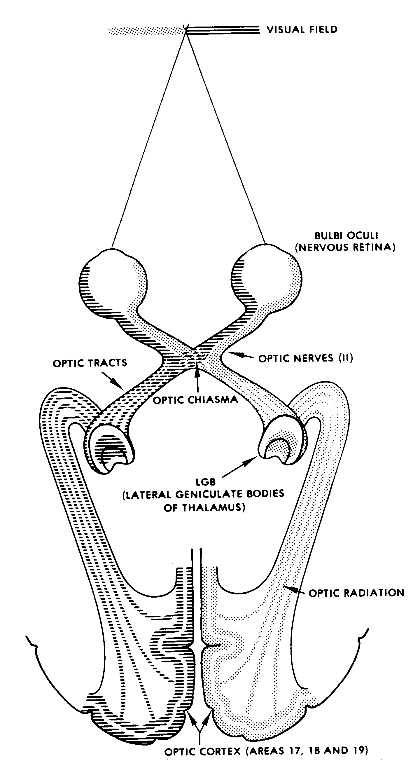
Figure 13-3. Scheme of visual input.
(1) Cones. The cones of the retina are for acute vision and also receive color information. The cones tend to be concentrated at the rear of the eyeball. The greatest concentration is within the macula lutea at the inner end of the focal axis (Figure 13-1).
(2) Rods. Light received by the rods is perceived in terms of black and white. The rods are sensitive to less intensive light than the cones. The rods are concentrated to the sides of the eyeball.
(3) Signal transmission. The stimulus from the photoreceptors (cones and rods) is transferred to the bipolar cells. In turn, the stimulus is transferred to the ganglion cells, the cells of the innermost layer of the retina. The axons of the ganglion cells converge to the back side of the eyeball. The axons leave the eyeball to become the optic nerve, surrounded by a dense FCT sheath. There are no photoreceptors in the circular area where the axons of the ganglion cells exit the eyeball; thus, this area is called the blind spot.
13-4. NERVOUS PATHWAYS FROM THE RETINAS
a. The two optic nerves enter the cranial cavity and join in a structure known as the optic chiasma. Leading from the optic chiasma on either side of the brainstem is the optic tract. In the optic chiasma, the axons from the nasal (medial) halves of the retinas cross to the opposite sides. Thus, the left optic tract contains all of the information from the left halves of the retinas (right visual field), and the right optic tract contains all of the information from the right halves of the retinas (left visual field).
b. The optic tracts carry this information to the LGB (lateral geniculate body) of the thalamus. From here, information is carried to the posterior medial portions (occipital lobes) of the cerebral cortex, where the information is perceived as conscious vision. Note that the right visual field is perceived within the left hemisphere, and the left visual field is perceived within the right hemisphere.
c. The LGB also sends information into the midbrainstem. This information is used to activate various visual reflexes.
13-5. FOCUSING OF THE LIGHT RAYS
a. The light rays, which enter the eyeball from the visual field, are focused to ensure acute vision. The majority of this focusing is accomplished by the permanently rounded cornea.
b. Fine adjustments of focusing, for acuteness of vision, are provided by the crystalline lens (biconvex lens). See Figure 13-4. This is particularly important when changing one's gaze between far and near objects.
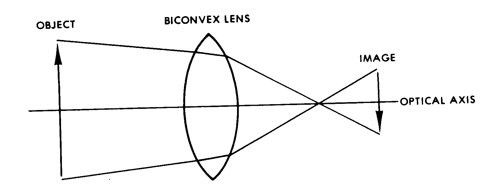
Figure 13-4. Bending of the light rays by a biconvex lens.
13-6. ACCOMMODATION
The additional focusing provided by the crystalline lens, mentioned above, is one of the processes involved in accommodation. Accommodation refers to the various adjustments made by the eye to see better at different distances.
a. The crystalline lens is kept in a flattened condition by the tension of the zonular fibers (zonule ligaments; fibers of the ciliary zonule) around its equator, or margin. Contraction of the ciliary muscle of the eyeball releases this tension and allows the elastic lens to become more rounded. Since the elasticity of the crystalline lens decreases with age, old people may find it very difficult to look at close objects.
b. A second process in accommodation is the constriction of the pupils. The diameter of the pupil (the hole in the middle of the iris) controls the amount of light that enters the eyeball. As a light source comes closer and closer, the intensity of the light increases greatly. Therefore, the pupils must be constricted to control the amount of light entering the eyeball as an object under view comes close to the individual.
c. A third process in accommodation is the convergence of the axes of the two eyeballs toward the midline. Since both eyes tend to focus on the same object (binocular vision), there is an angle between the two axes. As an object draws closer, the angle increases to enable the axes to still intersect the object.
13-7. EYE MOVEMENTS
a. Convergent and Conjugate Eye Movements. In a conjugate eye movement, both eyeballs move through an equal angle in one direction, such as right or left. In a convergent eye movement, both eyeballs turn toward the midline to focus upon a nearby object. In both cases, the movement of the left and right eyeballs is highly coordinated so that an object may be viewed by both eyes. Therefore, the object can be perceived within both cerebral hemispheres in a binocular fashion.
b. "Searching" and "Following" Eye Movements. "Searching" and "following" movements of the eyeball are also called, respectively, voluntary fixation movements and involuntary fixation movements. For the first type of movement, the eyeballs move in a searching pattern, without focusing on a particular object until it is located. Once an object is located, the eyeballs will continually fix on that object in a following-type motion.
c. Eye Movements During Reading. During reading of printed or written material, the eyeball demonstrates several physical characteristics. The amount of material that can be recognized at a given glance occupies a given width of a written line. Each glance is referred to as a fixation. During a fixation, the eyeball is essentially not moving, and each eyeball is oriented so that the image falls upon the macula lutea (the maximum receptive area). Reading is a series of motions in which the eyeballs fixate on a portion of the written line and then move very rapidly to the next portion.
d. Compensation for Head Movements (Vestibular Control of Eye Movements). Since the human body cannot be held absolutely still, the eyeballs must move in order to remain fixed upon an object. For this purpose, the eyeballs must be moved in the opposite direction and at the opposite speed of the movement of the head. This is accomplished by a delicate and complicated mechanism. This mechanism includes the motor neurons of the muscles of the eyeball and the vestibular nuclei of the hindbrain (responsible for balance and spatial orientation).
13-8. VISUAL REFLEXES
In the sense of vision, one consciously perceives the various objects being looked upon. In addition to this, there are a number of protective reactions to visual input--the visual reflexes.
a. When an unexpected visual stimulus occurs within the visual field, the individual's response will often include movement and other types of reaction. This is a part of the startle reflex.
b. When there is a change in the amount of light entering the eyeball, the size of the pupil will change. This is the pupillary reflex. The muscles of the iris automatically constrict or dilate to control the amount of light entering the eyeball.
c. In the blink reflex, the eyelids automatically move over the exterior surface of the eyeball. This reflex results in the automatic washing of the exterior surface of the eyeball with the lacrimal fluids. It also helps to keep the surface moist.
13-9. LACRIMAL APPARATUS
The eyeball is suspended in the orbit and faces outward. Helping to fill the orbit are a number of structures associated with the eyeball; these are the adnexa. Among these other structures is the lacrimal apparatus.
a. The lacrimal gland is located in the upper outer corner in front. Via small ducts, it secretes the lacrimal fluid into the space between the external surface of the eyeball and the upper eyelid.
b. The inner surface of the eyelids and the outer surface of the eyeball are covered by a continuous membrane known as the conjunctiva. The lacrimal fluid keeps the conjunctiva transparent. Also, with the blink reflex, the lacrimal fluid washes away any foreign particles that may be on the surface of the conjunctiva.
c. The free margins of the upper and lower eyelids have special oil glands. The oily secretion of these glands helps prevent the lacrimal fluid from escaping.
d. With the movement of the eyeball and the eyelids, the lacrimal fluid is gradually moved across the exterior surface of the eyeball to the medial inferior corner. Here, the lacrimal fluid is collected into a lacrimal sac, which drains into the nasal chamber by way of the nasolacrimal duct. Thus, the continuous production of lacrimal fluid is conserved by being recycled within the body.
Section III. THE SPECIAL SENSE OF HEARING (AUDITORY SENSE)
13-10. INTRODUCTION
If a medium is set into vibration within certain frequency limits (average between 25 cycles per second and 18,000 cycles per second), we have what is called a sound stimulus (Figure 13-5). The sensation of sound, of course, occurs only when these vibrations are interpreted by the cerebral cortex of the brain at the conscious level.
a. The human ear is the special sensory receptor for the sound stimulus. As the stimulus passes from the external medium (air, water, or a solid conductor of sound) to the actual receptor cells in the head, the vibrations are in the form of (1) airborne waves, (2) mechanical oscillations, and (3) fluid-borne pulses.
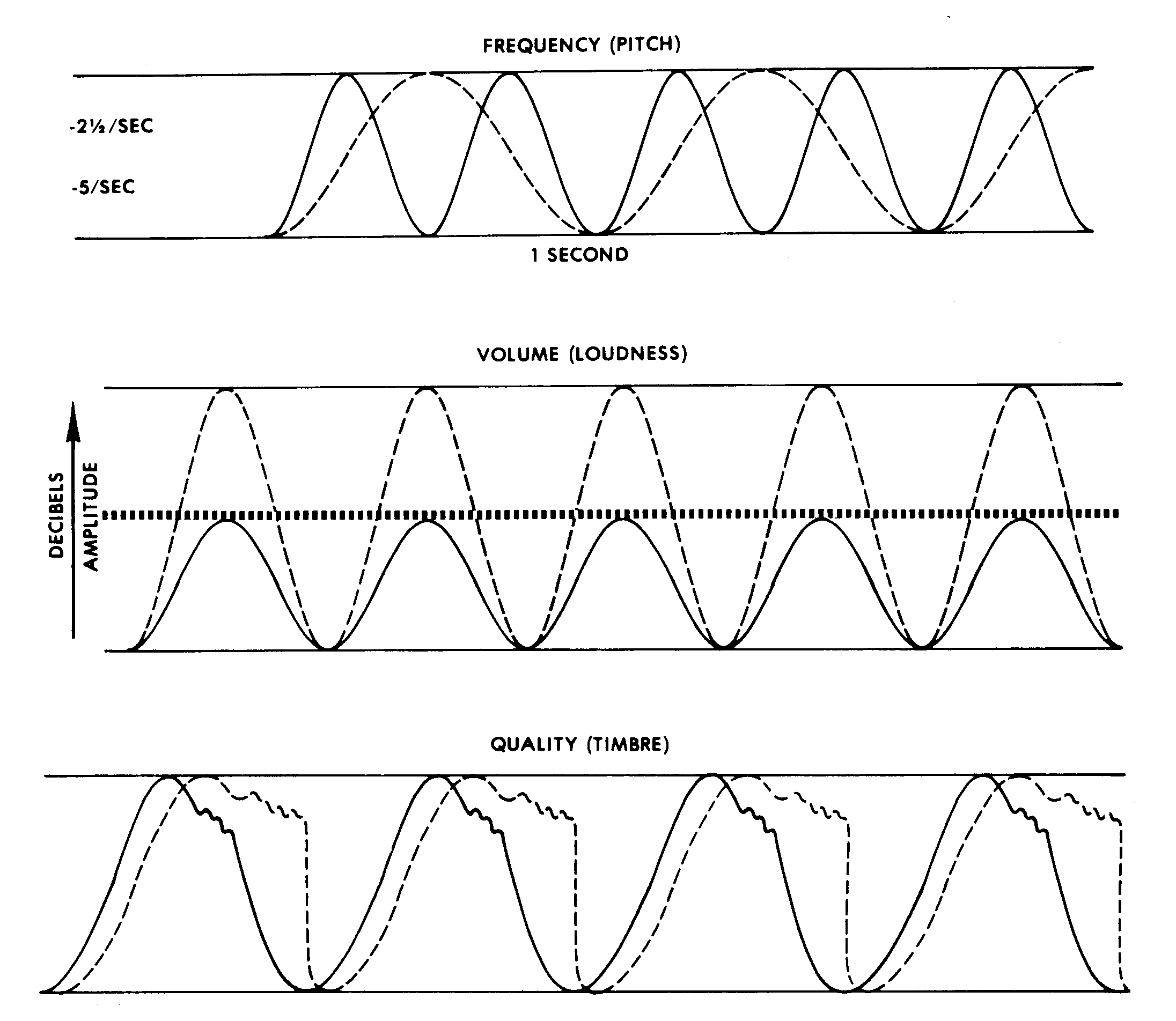
Figure 13-5. Characteristics of sound.
b. The ear (Figure 13-6) is organized in three major parts: external ear, middle ear, and internal (inner) ear. Each part aids in the transmission of the stimulus to the receptor cells.
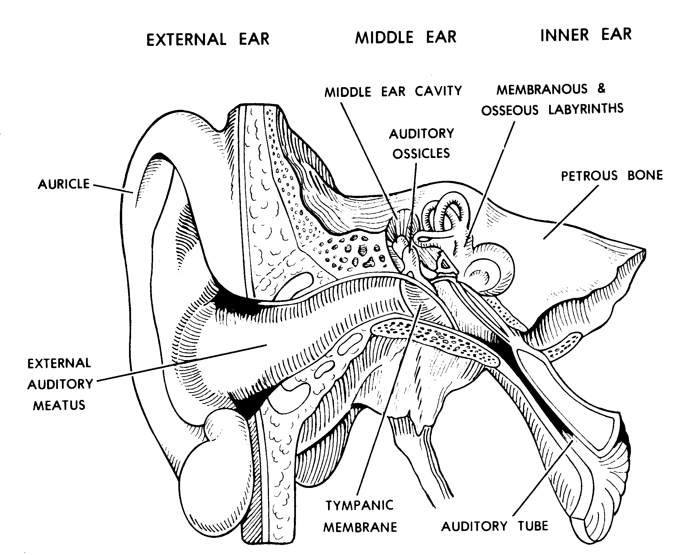
Figure 13-6. A frontal section of the human ear.
13-11. THE EXTERNAL EAR
The external ear begins with a funnel-like auricle. This auricle serves as a collector of the airborne waves and directs them into the external auditory meatus. At the inner end of this passage, the waves act upon the tympanic membrane (eardrum). The external auditory meatus is protected by a special substance called earwax (cerumen).
13-12. THE MIDDLE EAR
a. Tympanic Membrane. The tympanic membrane separates the middle and external ears. It is set into mechanical oscillation by the airborne waves from the outside.
b. Middle Ear Cavity. Within the petrous bone of the skull is the air-filled middle ear cavity.
(1) Function of the auditory tube. Due to the auditory tube, the air of the middle ear cavity is continuous with the air of the surrounding environment. The auditory tube opens into the lateral wall of the nasopharynx. Thus, the auditory tube serves to equalize the air pressures on the two sides of the tympanic membrane. If these two pressures become moderately unequal, there is greater tension upon the tympanic membrane; this reduces (dampens) mechanical oscillations of the membrane. Extreme pressure differences cause severe pain. The passage of the auditory tube into the nasopharynx opens when one swallows; therefore, the pressure differences are controlled somewhat by the swallowing reflex.
(2) Associated spaces. The middle ear cavity extends into the mastoid bone as the mastoid air cells. The relatively thin roof of the middle ear cavity separates the middle ear cavity from the middle cranial fossa.
c. Auditory Ossicles. There is a series of three small bones, the auditory ossicles, which traverse the space of the middle ear cavity from the external ear to the internal ear. The auditory ossicles function as a unit.
(1) The first ossicle, the malleus, has a long arm embedded in the tympanic membrane. Therefore, when the tympanic membrane is set into mechanical oscillation, the malleus is also set into mechanical oscillation.
(2) The second ossicle is the incus. Its relationship to the malleus produces a leverage system which amplifies the mechanical oscillations received through the malleus.
(3) The third ossicle, the stapes, articulates with the end of the arm of the incus. The foot plate of the stapes fills the oval (vestibular) window.
d. Auditory Muscles. The auditory muscles are a pair of muscles associated with the auditory ossicles. They are named the tensor tympani muscle and the stapedius muscle. The auditory muscles help to control the intensity of the mechanical oscillations within the ossicles.
13-13. THE INTERNAL EAR
a. Transmission of the Sound Stimulus. The foot plate of the stapes fills the oval (vestibular) window, which opens to the vestibule of the internal ear (Figure 13-7A). As the ossicles oscillate mechanically, the stapes acts like a plunger against the oval window. The vestibule is filled with a fluid, the perilymph. These mechanical, plunger-like actions of the stapes impart pressure pulses to the perilymph.
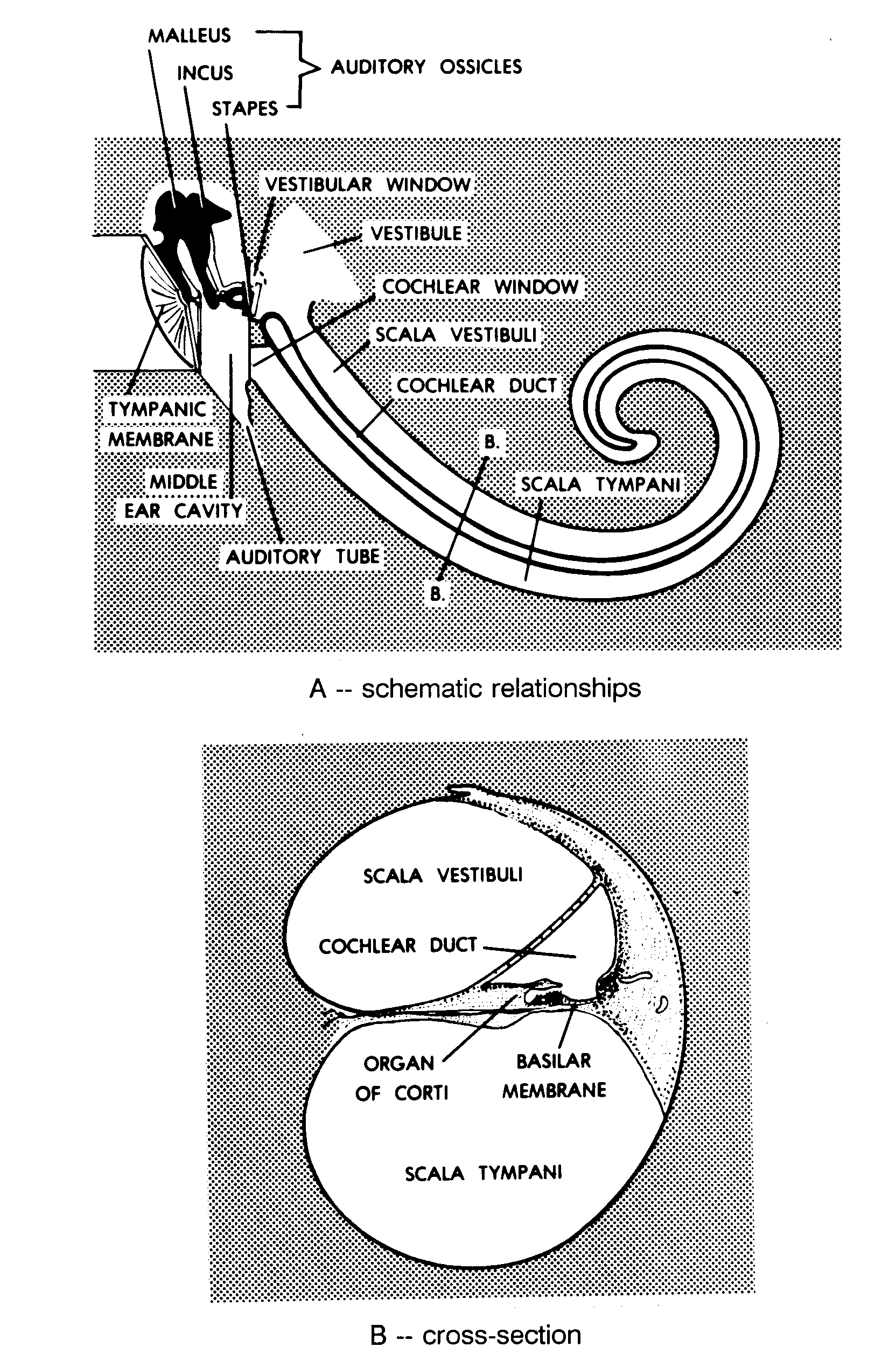
Figure 13-7. Diagram of the scalae.
b. Organization of the Internal Ear. The internal ear is essentially a membranous labyrinth suspended within the cavity of the bony (osseous) labyrinth of the petrous bone (Figure 13-8). The membranous labyrinth is filled with a fluid, the endolymph. Between the membranous labyrinth and the bony labyrinth is the perilymph.
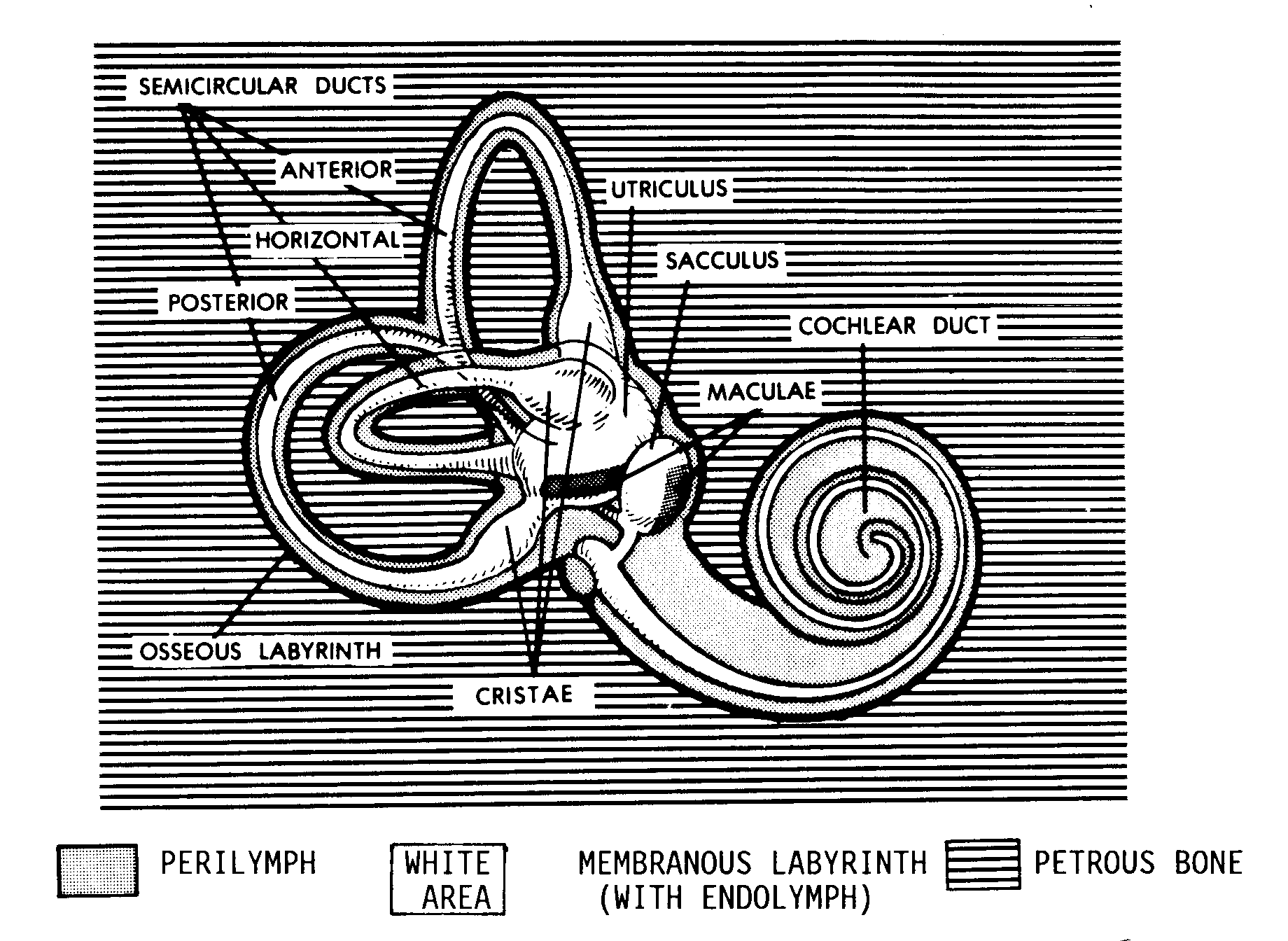
Figure 13-8. The labyrinths of the internal ear.
c. The Cochlea. The cochlea is a spiral structure associated with hearing. Its outer boundaries are formed by the snail-shaped portion of the bony labyrinth. The extensions of the bony labyrinth into the cochlea are called the scala vestibuli and the scala tympani (Figure 13-7B). These extensions are filled with perilymph.
(1) Basilar membrane (Figure 13-7B). The basilar membrane forms the floor of the cochlear duct, the spiral portion of the membranous labyrinth. The basilar membrane is made up of transverse fibers. Each fiber is of a different length, and the lengths increase from one end to the other. Thus, the basilar membrane is constructed similarly to a harp or piano. Acting like the strings of the instrument, the individual fibers mechanically vibrate in response to specific frequencies of pulses in the perilymph. Thus, each vibration frequency of the sound stimulus affects a specific location of the basilar membrane.
(2) Organ of Corti. Located upon the basilar membrane is the organ of Corti. The organ of Corti is made up of hair cells. When the basilar membrane vibrates, the hair cells are mechanically deformed so that the associated neuron is stimulated.
13-14. NERVOUS PATHWAYS FOR HEARING
The neuron (associated with the hair cells of the organ of Corti) then carries the sound stimulus to the hindbrainstem. Via a special series of connections, the signal ultimately reaches Brodmann's area number 41, on the upper surface of the temporal lobe (see para 12-36). Here, the stimulus is perceived as the special sense of sound. It is interesting to note that speech in humans is primarily localized in the left cerebral hemisphere, while musical (rhythmic) sounds tend to be located in the right cerebral hemisphere.
Section IV. THE SPECIAL SENSE OF EQUILIBRIUM, THE GENERAL BODY
SENSE, AND POSTURAL REFLEXES
13-15. INTRODUCTION
a. The human body is composed of a series of linkages, block on top of block. These blocks can be arranged in a multitude of patterns called postures. In order to produce and control these postures, the human brain utilizes a great number of continuous inputs telling the brain the instantaneous condition of the body posture. Overall, we refer to this process as the general body sense.
b. The internal ear provides one of the input systems for the general body sense. The internal ear responds to gravitational forces, of which there are two kinds--static and kinetic (in motion). Of the kinetic stimuli, the motion may be in a straight line (linear) or angular (curvilinear).
13-16. THE MACULAE
The membranous labyrinth of the internal ear has two sac-like parts--the sacculus and the utriculus. On the wall of each of these sacs is a collection of hair cells known as the macula (plural: maculae). The hairs of these hair cells move in response to gravitational forces, both static and linear kinetic. The maculae are particularly sensitive to small changes in the orientation of the head from an upright position. Thus, the maculae are very important in maintaining a standing or upright position.
13-17. THE SEMICIRCULAR DUCTS
a. In addition, three tubular structures are associated with the utriculus. The circle of each of these semicircular ducts is completed by the cavity of the utriculus. At one end of each semicircular duct is a crista, a ridge of hair cells across the axis of the duct.
b. When a jet takes off, a passenger tends to remain in place at first and can feel the resulting pressure of the seat against his back. Also, when the jet is no longer accelerating, the passenger can feel that the pressure of the seat against his back has returned to normal.
c. Likewise, in the appropriate semicircular duct, the endolymph ("passenger") tends to remain in place early during an acceleration. Because the duct ("seat") itself is moving with the body ("jet"), the hairs of the crista are affected by the change in movement. Later, when acceleration stops, the effect upon the hairs of the crista is also registered.
d. However, the cristae of the semicircular ducts detect rotation of the head (angular acceleration and angular velocity). Linear acceleration, as with our example of the passenger and the jet, is detected primarily by the maculae, discussed above.
13-18. RESULTING INPUTS FOR THE SPECIAL SENSE OF EQUILIBRIUM
The combined inputs from the maculae of the sacs and the cristae of the semicircular ducts provide continuous, instantaneous information about the specific location and posture of the head in relationship to the center of gravity of the earth. These inputs are transmitted by the vestibular neurons to the hindbrainstem.
13-19. INPUTS FOR THE GENERAL BODY SENSE
In addition to the inputs from the membranous labyrinth, various other inputs are used to continuously monitor the second-to-second posture of the human body.
a. We have already examined the proprioceptive sense, which monitors the condition of the muscles of the body.
b. Various other receptors are associated with the joint capsules, the integument, etc. They indicate the precise degree of bending present in the body.
c. A very important body sense is vision. Even when other inputs are lacking, if an individual can see his feet, he may still be able to stand and move.
13-20. POSTURAL REFLEXES
To automatically control the posture, the human nervous system has a number of special reflexes. These reflexes are coordinated through the cerebellum.
a. The head and neck tonic reflexes orient the upper torso in relationship to the head.
b. Another set of reflexes does likewise for the body in general. The righting reflexes come into play when the body falls out of balance or equilibrium.
c. A special set of reflexes connects the vestibular apparatus to the extraocular muscles of the eyeball. This was discussed earlier in the section on the special sense of the vision.
Section V. THE SPECIAL SENSE OF SMELL (OLFACTION)
13-21. SENSORY RECEPTORS
Molecules of various materials are dispersed (spread) throughout the air we breathe. A special olfactory epithelium is located in the upper recesses of the nasal chambers in the head. Special hair cells in the olfactory epithelium are called chemoreceptors, because they receive these molecules in the air.
13-22. OLFACTORY SENSORY PATHWAY
The information received by the olfactory hair cells is transmitted by way of the olfactory nerves (cranial nerves I). It passes through these nerves to the olfactory bulbs and then into the opposite cerebral hemisphere. Here, the information becomes the sensation of smell.
Section VI. THE SPECIAL SENSE OF TASTE (GUSTATION)
13-23. SENSORY RECEPTORS
Molecules of various materials are also dispersed or dissolved in the fluids (saliva) of the mouth. These molecules are from the food ingested (taken in). Organs known as taste buds are scattered over the tongue and the rear of the mouth. Special hair cells in the taste buds are chemoreceptors to react to these molecules.
13-24. SENSORY PATHWAY
The information received by the hair cells of the taste buds is transmitted to the opposite side of the brain by way of three cranial nerves (VII, IX, and X). This information is interpreted by the cerebral hemispheres as the sensation of taste.